Overview
This human FFPE clear cell renal cell carcinoma (ccRCC) dataset was generated following the Xenium In Situ Gene and Protein Expression with Cell Segmentation Staining workflow. The Xenium Human Multi-Tissue and Cancer Panel, supplemented with custom genes, was used to profile gene expression. The 27-plex Cell Typing & Functional State Marker Set was used to profile protein expression (immune cell types, functional states, and tissue microenvironment context).
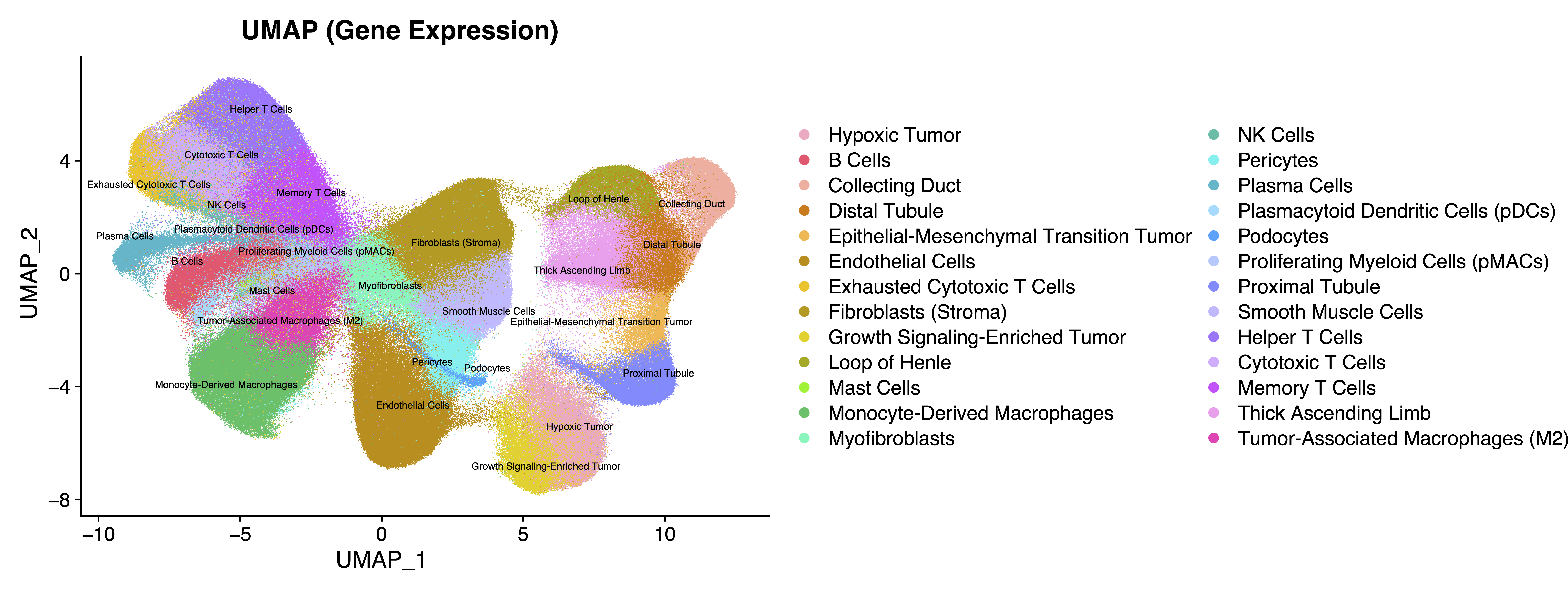
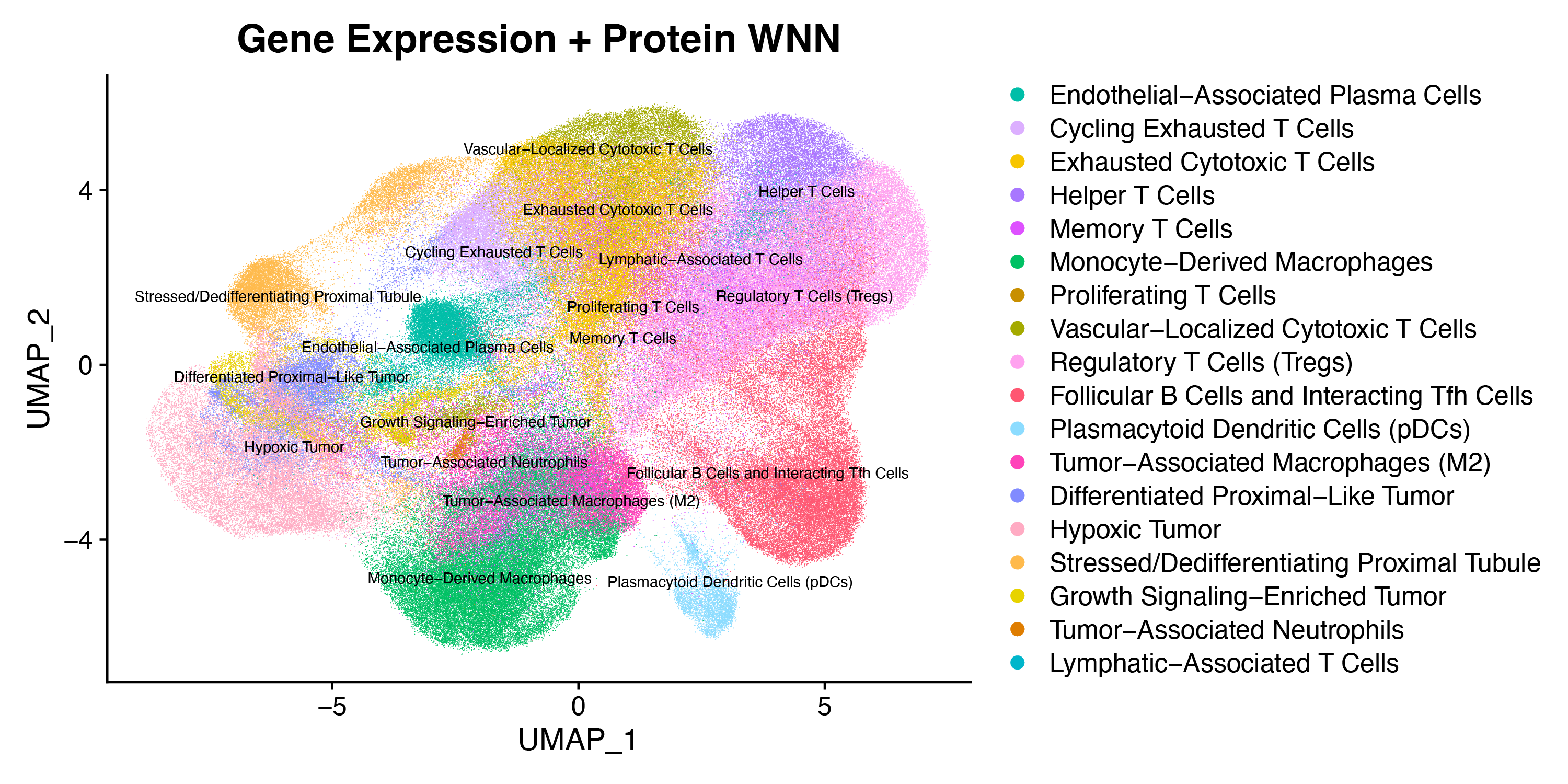
This multiomic dataset was analyzed using a weighted-nearest neighbor (WNN) clustering approach1 to integrate RNA and protein data (see custom cell groups description for details). The method captured maximum biological variability, allowing us to identify spatially heterogeneous expression across tumor regions. Key markers of interest include RNA targets (SOSTDC1, DPP4, CCND1, ADAM28, AIF1, APOL1, CD47, CXCL2, CXCL6, CXCL8, FSTL3, S100A, S100B) and proteins (β-catenin, PTEN), all of which are linked to tumor progression and vary by region. The annotated UMAPs below illustrate enhanced biological insight gained by integrating RNA and protein data, including possible cellular localization, as well as multiple stages and forms of tumor cell types.
UMAP showing annotated cell types in ccRCC RNA gene expression data:
UMAP showing immune cell subset clustering from WNN integration of RNA and protein data:
How to view data
Interactively explore data with Xenium Explorer by downloading the Xenium Output Bundle (or Xenium Explorer subset) file. The subset bundle contains the experiment.xenium, gene_panel.json, morphology_focus/ directory of multi-file OME-TIFF files, analysis_summary.html, cells.zarr.zip, cell_feature_matrix.zarr.zip, transcripts.zarr.zip, and analysis.zarr.zip files.
See the Getting Started with Xenium Explorer page for more details. Follow these instructions to view the post-Xenium H&E image or image alignment file in Xenium Explorer.
Biomaterials
FFPE tissue blocks were purchased from AcePix Biosciences (kidney cancer, Clear Cell Renal Cell Carcinoma).
Tissue preparation
Tissue was prepared following the Xenium In Situ for FFPE - Tissue Preparation Handbook (CG000578).
Probe hybridization, washing, ligation, amplification, and cell segmentation staining were performed following the Xenium In Situ Gene and Protein Expression with Cell Segmentation Staining User Guide (CG000819).
Post-instrument processing followed the Xenium In Situ Gene Expression - Post-Xenium Analyzer H&E Staining Demonstrated Protocol (CG000613). The image was tiled, stitched, and acquired on an Olympus VS200 Slide Scanner. The image was registered following this tutorial.
Gene panel
The Xenium Human Multi-Tissue and Cancer Panel (377 genes) was pre-designed by 10x Genomics. The panel design was informed using single cell RNA sequencing data curated and reprocessed for standardization by the Human Protein Atlas. Genes were chosen to accurately type cells and identify select immune, proliferation, and tumor markers, in human breast, lung (healthy, cancer), skin, liver, colon, kidney, and heart.
We included 100 custom RNA targets covering: (1) genes encoding the proteins on the Xenium Protein subpanels, (2) kidney cell types not present in the pre-designed gene panel, and (3) genes implicated in renal carcinoma pathology.
Protein panel
The protein panel for this dataset consisted of all six Xenium Protein subpanels. It also included separately imaged cell segmentation (Xenium Multi-Tissue Stain Mix) markers (E-cadherin, Vimentin, alphaSMA, CD45), which are labeled "Cellular Localization" markers in outputs.
Xenium Analyzer
The instrument run was performed following the Xenium Analyzer User Guide (CG000584). The on-instrument analysis was run with Xenium Onboard Analysis v4.0.
| Metric | ccRCC |
|---|---|
| Median transcripts per cell | 54 |
| Cells detected | 690,322 |
| Nuclear transcripts per 100 µm² | 182.4 |
| High quality decoded transcripts detected | 65,480,008 |
| Region area (µm²) | 114,121,391.1 |
Custom Cell Groups
The following approach was used to integrate and annotate multiomic data from a single renal tumor section in Seurat.
- Cells were retained if they had non-zero RNA counts and total protein within ±4 SD of the log1p mean (z-score filter). Cells at the tissue edge with low quality and spatially inconsistent cell type annotations were manually removed (n = 8,927 cells), yielding 639,619 final cells for analysis.
- Gene expression was log-normalized, scaled, and reduced by PCA, followed by UMAP and Leiden clustering (18 PCs, k = 15, resolution = 0.6).
- Protein counts were independently normalized by centered log-ratio (CLR) transformation, scaled, and clustered (15 PCs, resolution = 0.6).
Major compartments were annotated from canonical markers (Seurat FindAllMarkers, Wilcoxon test, min.pct = 0.15, log2FC ≥ 0.25) and refined through iterative sub-clustering of the epithelial/tumor and immune compartments (SCTransform re-normalization within each subset). To integrate modalities, RNA and protein data were jointly modeled by weighted nearest-neighbor (WNN) analysis (FindMultiModalNeighbors) only on immune or tumor cell types (n = 15 cell types), combining RNA (15 dimensions) and protein (10 dimensions) PCA embeddings, followed by WNN-UMAP and Leiden clustering (resolution = 0.3). Restricting WNN analysis to immune and tumor cells yielded the most focused results for this analysis based on the Xenium Protein subpanels used in this experiment (this is not a universal analysis step for all samples or experiments). Final cell type labels were assigned by manual review of cluster markers in both modalities.
References
1 Hao Y, et al. Integrated analysis of multimodal single-cell data. Cell 184: 3573–3587.e29, 2021.