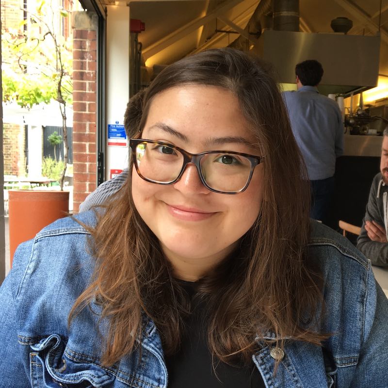
Why I chose spatial: New pathways into clinical translational research
In Part 1 of the “Why choose spatial” series, we shared insights from our tissue and spatial biology experts regarding key considerations before choosing a solution. In Part 2, researchers share their stories of why they adopted spatial tools into their workflows and what spatial analysis has done to advance their research.
Why did Choi Hongyoon, MD, PhD, Assistant Professor at Seoul National University Hospital, choose spatial? For him, it was a logical solution. With previous research in how imaging data could be extended with molecular information for clinical purposes, Dr. Choi was interested in combining nuclear imaging and omics techniques to study the tumor microenvironment. But, while both methods provide invaluable insights, he knew they would be even stronger together. He just needed a way to link the two. That’s why he initially turned to spatial transcriptomics. It was the perfect bridge to connect these data streams—but that wasn’t the end of the story.
Sometimes, you don’t just cross the bridge to get to the other side. A particularly good view might stop you in your tracks right in the middle. Dr. Choi, his spatial bridge in place, took in the view and saw the possibilities—how the integration of spatial transcriptomics with imaging and other techniques could create myriad potential new pathways for translational research.
So, for Dr. Choi, what was originally one reason to choose spatial has now become many. Read our Q&A with Dr. Choi to find out how spatial biology is helping him find innovative new ways to further his clinical translational research.

Could you please describe your current research projects?
I am conducting a project to find new targets for characterizing the tumor microenvironment and develop applications by integrative analysis of imaging and spatial transcriptomics. In particular, through collaboration with the lung cancer team, based on human lung cancer data, spatial transcriptomics is integrated with other data (imaging and genomic) to analyze tumor microenvironment and cancer heterogeneity. During this project, I and collaborators focus on how to extend and translate spatial molecular information into clinical imaging and therapy.
How did you become interested in studying your field?
While carrying out clinical jobs as a medical doctor and R/D projects in hospitals, I watched data and AI rapidly innovate in the biomedical field. In the field of medical imaging, I made several applications through deep learning, and I wanted to turn this to a more innovative direction. So, I thought that it could contribute not only to imaging obtained clinically, but also to the development of new biomarkers and therapeutics. And I thought that this application could be started from “omics” data.
My other research interest regarding clinical work includes in vivo targeting of specific molecules for developing imaging and therapeutics, which is one of the goals of nuclear medicine fields in terms of translational research. In this process, I have taken new approaches by using AI for image analysis and integrating that with molecular information obtained by omics studies. The study started on how clinically used molecular imaging (e.g., PET) can be clinically extended by combining it with omics information (1). In terms of the spatial scale, an unmet need in this process was the lack of a link between imaging that can be used clinically and omics information based on biological studies. Thus, I became interested in spatial transcriptomics as a solution to link them.
What led you to consider using spatial technology to answer your research questions?
As we began research in this way, we realized that spatial molecular information has infinite scalability. In particular, when this spatial molecular information is combined with other data (in my case, imaging), the idea that it can be used in various ways—such as discovering new targets for drugs, verifying drug target delivery as a preclinical study, and imaging biomarker technology for companion diagnostics of new drugs—is spreading. As such, we are working hard on the R&D, focusing on the new translational applications of spatial omics.
What were the most important findings enabled by spatial technology from your studies?
Currently, we are developing solutions one by one in order to continue to find our answers: imaging biomarkers and new molecular targets for clinically applicable imaging and therapeutics.
First of all, although it is an early form, we have proposed an initial technique that can be interpreted by combining imaging and spatial transcriptomics (2). This could be useful in that it can be spatially interpreted in various ways by placing H&E or other images (e.g., protein expression, ex vivo drug distributions) on the Visium data. In particular, the method includes an approach that extracts image features through deep learning and analyzes them in relation to molecular information. We think this will be extended to combine different kinds of imaging and spatial transcriptomics to find new molecular information within spatial biology.
Another approach was developed with the motivation that it was difficult to interpret the cell in spatial transcriptomics analysis. Therefore, a new method combined with the cell type of single cell RNA-seq was needed. The CellDART technology we made utilizes a deep learning–based technology called domain adaptation, and provides a fast and accurate interpretation of spatial data to the cell-type level. It accurately maps the cell types to the spatial transcriptomic data (3).
How has taking a spatial approach informed your next research questions or follow up experiments?
We continue to converge this data to develop solutions for clinical translational applications. In this process, targets specific to cancer or specific cell types were found through single cell and spatial transcriptomics. Therefore, we are promoting and collaborating research and development in the form of target-based drug development (e.g., radiolabeled therapeutics, radioligand therapy) through various knowledge-based filters. In addition, spatial transcriptomics is applied and utilized as a platform for developing AI-based imaging biomarkers through combination with images.
Judging that there is sufficient scalability in this process, I and my colleagues founded Portrai, Inc. that can contribute to clinically applicable biomarker and drug development based on spatial technology, and we are eager to continue R&D to make practical clinical application solutions.
Why was Visium Spatial Gene Expression from 10x Genomics essential for your findings?
To the fundamental question of how to find key molecular information that can be translated to a clinical scale (i.e., whole body and systemic scale) in a lot of molecular information, the important point is that Visium can provide that link because of “intermediate” spatial scale.
What led you to choose Visium over other spatial technologies?
I believe that innovative new drugs and treatment concepts and AI-based biomarkers that are starting to be used in current clinical practice will be innovated through “data.” By providing the most valuable information at once at these points with intermediate scale that can be interpreted and imaged in the clinical point, Visium Spatial Gene Expression will become a driving force for data-based drug and biomarker development beyond hypothesis-based development. And as a translational researcher and a clinician, I would like to contribute to creating new imaging methods and therapeutics based on spatial technology for specific solutions and realizations of these data-based innovations.
References:
- Park C, et al. Tumor immune profiles noninvasively estimated by FDG PET with deep learning correlate with immunotherapy response in lung adenocarcinoma. Theranostics 10(23): 10838 (2020). doi: 10.7150/thno.50283
- Bae S, et al. Discovery of molecular features underlying the morphological landscape by integrating spatial transcriptomic data with deep features of tissue images. Nucleic Acids Res 49(10): e55-e55 (2021). doi: 10.1093/nar/gkab095
- Bae S, et al. CellDART: cell type inference by domain adaptation of single-cell and spatial transcriptomic data. Nucleic Acids Res 50(10): e57-e57 (2022). doi: 10.1093/nar/gkac084
