The Neuroscience Journal Club: Connecting you to the latest neuro research
Imagine this: you’re sitting in your department’s journal club, discussing an exciting new publication. You come across an interesting decision. Maybe the authors used FACS when you might’ve used MACS. Or, maybe they decided to analyze nuclei, when you’d have chosen whole cells.
Why did the authors make that choice? What led them to this experiment in the first place? What’s next? The possibilities are endless, but so are the unknowns.
It’s easy to imagine, right? Journal clubs are critical for keeping up with the latest discoveries in your field, and they can often serve as forums for valuable discussions with your peers. But there are times when it would be nice to ask the authors your questions directly and get the answers.
That’s where the 10x Genomics Neuroscience Journal Club comes in. Our new quarterly webinar series brings you the authors of some of the most conversation-worthy new neuroscience publications, so you can get the answers to your questions—live.
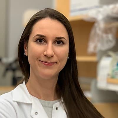
Want a preview? Our first meeting of the Neuroscience Journal Club featured Galina Popova, PhD, a postdoctoral researcher in the Nowakowski Lab at UCSF, as she discussed her recent Cell Stem Cell publication. During the meeting, she walked us through her research, describing how she and her colleagues used single cell technologies from 10x Genomics to study microglial function across models.

The Microglia Report Card: Choosing the right experimental model
Using single cell RNA-sequencing (scRNA-seq), the team analyzed microglia across common experimental models, including cultured primary and pluripotent stem cell–derived microglia, identifying five molecularly unique clusters. Each microglia subtype was given a score representing its top 15 differentially expressed markers as well as its dividing cell signature. The scores were then used to create a “microglia report card,” that Popova and her colleagues used to compare microglial response across models, perturbations, and disease states, as well as key transcriptional programs of developing microglia in vitro.
Armed with this critical information, Popova and her team hypothesized that a 3D organoid environment would preserve homeostatic signatures in microglia. Transplanting human microglia into cerebral organoids, they used scRNA-seq to characterize key transcriptional programs of developing microglia in vitro. They discovered that microglia play an important role in the developing human brain, inducing transcriptional changes in neural stem cells.
Throughout the presentation, attendees were encouraged to submit their questions on the experiment, and Popova did her best to answer them all. Keep reading to see some of her conversation with neuroscience 10x-pert and Associate Director of Market Development Kelly Miller, PhD. Then, check out the full on-demand recording here.
Sample Prep
Can you briefly describe the process of isolating the microglia or purifying them from the organoids in order to process them for the single cell RNA-seq?
Galina Popova: Yes, from the protocol standpoint, that's also a very good question because it took me several attempts to collect enough microglia cells. Here, I would take organoids with microglia that I knew had already transplanted there, and I would start with 15 to 20 organoids. I dissociated them enzymatically. After that, I purified the microglia using CD11b magnetic beads from MACS. And with that, I was able to get enough cells for single cell profiling.
Can you comment on how many cells you ended up using for the single cell sequencing? What's recommended, especially given some of the kind of low RNA content of microglia and sometimes low expression of certain microglial genes like cytokines and things like that?
Galina Popova: No, I think actually they weren't particularly low RNA-content cells. Here, we used roughly 20,000 cells. I think that amount of cells is good enough to answer our question—how well different scores that were identified in primary microglia are preserved in, we call them 3D-cultured, microglia? And also 20,000 cells was a good amount to uncover some of the novel signatures that we didn't see in the in vivo developing microglia. So, yeah, it was a little bit challenging to get enough microglia cells for profiling, but, once we had our single cell RNA sequencing results, that wasn't an issue, I would say. And when we look at those different projected scores on the microglial UMAP space, again, we found preservation of homeostatic and ATM signatures, but somewhat in contrast to the mouse in vivo–cultured microglia. The separation of those signatures was a little bit more muted in organoids. But one of the most prominent scores that we got in microglia from the organoids was the cytokine-associated signature. That was really strong, and it was pretty much at the level of what we saw with in vivo microglia in prenatal brain.
How do different microglia purification strategies influence gene expression profiles?
Galina Popova: I also had this question in mind when we were working on this data. At some point, I generated a dataset from MACS-purified microglia from a prenatal human brain. And, while I'm not showing it in this paper, we compared it to cells that we extracted from the total the BICCN dataset that didn't have any antibody-based purification of microglia. We also recovered the same signatures in both datasets. So, I don't think we introduce— I don't think we, let's say, select some population of microglia that we would not be able to select if we used the whole brain. So I think the representation of different microglial subtypes is preserved with at least CD11b MACS purification.
When you use dissociated prenatal human tissue, do you use whole brain or do you have some reproducible method for a targeted dissection of the tissue?
Galina Popova: I primarily worked with cortical microglia, so the majority of the data would be cortical in this regard. It's a really good question, given that region-associated signatures exist among different microglia cell types.
How long did you incubate [iPSC-derived microglia] cells in the mouse brain prior to your single cell RNA-seq experiment?
Galina Popova: They “culture,” or incubate, microglia in the mouse brain for, I think, two weeks. It's done by injecting iMG [induced microglia-like] cells in a mouse brain and then, after a certain amount of time, they profile the human-specific microglia cells.
Have you tried using Dynabeads for your microglia isolation?
Galina Popova: Here everything was done on magnetically activated sorting, MACS, with CD11b beads. I think that's what they meant by Dynabeads. But, yes, all the microglia that I worked with were obtained through CD11b magnetic beads. Not FACS.
Was your tissue dissociation enzymatic or manual?
Galina Popova: It was enzymatic. Yes, it was trypsin at 37 degrees. So, some amount of heat-activated ex vivo activation signatures are expected.
Experimental considerations
Did all of the samples come from the same sex, as there have been some reports in the literature of gender-related microglial differences?
Galina Popova: We were agnostic to the sex of the samples here. Based on the BICCN dataset that we used to benchmark our models, all clusters were represented across different sexes, and there were some subtle differences between different sexes. But the majority of the scores or, shall I say, all of the scores that we found and that we're describing here were equally represented in both conditions.
What is the efficiency of integration? Would you need organoids to be a certain size to have a better integration?
Galina Popova: Yes, it's a good question. I didn't really compare the effect of size on the microglial transplantation. I did notice, somewhat anecdotally, that microglia don't transplant as well into the older organs. My explanation for that would be that the neural progenitor cells provide some of those trophic molecules that are necessary for microglial survival, and, over time, you see less and less of those neural progenitor cells in the organoids. So I think microglia actually kind of use both progenitors and more mature neurons for that trophic support.
The efficiency of immigration, it's not it's not awfully high. I usually transplant roughly 100,000 microglia cells per organoid, and this one is a section of roughly 20 microns. So, I get maybe thousands of cells per organoid, and not all microglia would find their way into the organoids and survive. But the ones that do, they become ramified and they start playing some of the roles that are expected of microglia, including, for example, phagocytic cups.
How long do the microglia survive in the organoids?
Galina Popova: It's interesting that, over time, the numbers go down, and I can't really explain why, exactly, because microglia need several trophic factors in order to survive and be well. And based on the 10x [Genomics] experiments that we did on the organoids, different cell types in the organoids should provide all of these trophic factors and molecule cues. In short, usually five weeks is what is reasonable to expect for microglial survival.
Have you tried any single nuc-seq with these organoids? Do you know if microglia show up well with nuc-seq, which would allow for microglia to be profiled potentially from snap frozen archived samples?
Galina Popova: I personally have not tried it. Several months ago, there was a paper that compared nuc-seq of microglia and whole cell of microglia, and they claim that nuc-seq does not preserve our cytokine-associated signature that well. So, I think you do lose at least some signatures if you remove all the cytoplasm from your data analysis. So for us, since the cytokine-associated microglia was one of the most prominent signatures, I think it would make a big difference.
Given that most cell types derived from pluripotent cell sources are functionally immature, how long in culture or transplant after the start of differentiation do you think is required for the iMGs to become functional?
Galina Popova: I guess it refers to culture in the organoids. Microglia are really highly dynamic, so one study found that if you extract microglia cells from the brain and you throw them on a 2D dish, you start seeing transcriptional differences as early as six hours. Given how highly dynamic the cells are, I would assume that it would not take too long for the microglia to mature under the influence of different brain cell-type populations. I would say between one to two weeks is probably the sweet spot to both induce maturation and still have the high number of iMG cells surviving, transplanted, and kind of being in this steady state in the organoids.
Findings
I know you also cited Marsh et al. in this publication. So in regard to that, the signatures that they published—or changes resulting from the enzymes—did you see any of that? Did you see something similar?
Galina Popova: We definitely saw a lot of that in our primary microglia. And again, these come from whole brain tissue that was dissociated for the 10x [Genomics] experiment. In this one, it's cluster 3, or ex vivo activation, we had a really robust signature in those cells. When we moved to different models, for example, primary cultured microglia, I cultured it for several days, and then, since it was just a 2D culture, removing them from the dish was much faster than using the whole brain. So, here, our ex vivo activation signature was much lower, simply because the processing time was much shorter for 2D-cultured microglia.
How do different scores compare, and which models are better or worse?
Galina Popova: So when we compared different scores across our models, one interesting observation was in this cytokine-associated microglia signature that I just mentioned in a previous slide. We saw the highest cytokine-associated gene expression in the primary human brain and also in microglia that were cultured inside of the organoids. And to me, it was interesting because these are two different models or conditions where microglia are surrounded by human cell types representing normal developing brain, and the same signature was more muted when microglia were cultured, or transplanted, in vivo mouse brain and when they were cultured without any other human cell populations. So, one interpretation here is that different cues that come from human-specific brain cells are enough to induce the cytokine signature, and the mouse brain cells may not be enough to actually induce this cytokine profile. In terms of the homeostatic signature, we found that, understandably, primary is the best to keep microglia nice and homeostatic, followed by the mouse brain, and then the organoid environment is still better than culturing microglia cells on their own.
Have you looked at all at microglia–astrocyte interaction?
Galina Popova: We didn't really focus on any cell–cell interactions per se. We decided to focus a little bit more on neural progenitor cells, first, because, in the developing brain, we found that these interactions are the most enriched and, second, astrocytes in organoids mature a little bit later, so it would take a slightly different timescale. So, the short answer to that is, no, not really.
Did you compare the organoid-resident microglial transcriptomic profile with that of brain-resident microglia? Do you know how the other cells in the organoid, such as radial glia or neurons, compared to their native state in the brain?
Galina Popova: Yes, the first part of the question—how microglia transcriptomics compare to brain-resident microglia. Gosselin et al., they looked— At first, I think the majority was in bulk, and they looked at a lot of mouse brain or adult human brains. So, we compared our prenatal microglia to in vivo developing human microglia. And I think it makes for a more fair comparison, including comparing across the same transcriptional single cell platform—10x [Genomics].
Comparing different organoid cell types, there are some other papers that actually looked at that, including some of the papers that we cite in ours. And the short answer—they represent, but not fully represent, what's going on in the in vivo human brain.
Did any of the microglial clusters that you identified resemble the disease-associated microglia (DAM) signatures?
Galina Popova: Yes, absolutely. Microglia are really context-specific, and DAM, originally found in Alzheimer's disease and named after the disease, were later found in developing microglia. I'm not saying that these are exactly the same cells because certain gene profiles are actually different between disease-associated microglia and developmental substate or subtype. But some of the genes are really similar, which makes me think that this population— I think, in terms of naming, we should be switching more to the functional association of different types instead of naming them based on the condition where we first found them.
Kelly Miller: Yes, I was going to say, this is very reminiscent of the M1/M2 macrophages, and we tried for a long time to move away from that, and now we have kind of a reversion to all of these new descriptors. So, yeah, I agree.
Galina Popova: Yes, and, even in this paper, we have this mix of abbreviated names for microglia, but we also try to associate them with individual gene profiles that are common across each subtype or each subcluster. But, yes, I think better and better nomenclature for substates will be really important for understanding and moving away from [the idea] that these are bad microglia or good microglia.
We want to give a big thank you to Dr. Popova, for sharing her research and taking the time to answer so many of our questions! Her presentation is now available to watch on-demand. And make sure to keep an eye out for our next Neuroscience Journal Club meeting, coming soon.
