#AACR18 Day 2 - Single Cell Genomics: Addressing Challenges in Cancer Research
Today, AACR 2018 was all about single cell genomics! My day started bright and early with the Plenary Session, "Elucidating the Complexities of Cancer," chaired by John Carpten, USC Keck School of Medicine. It was a great way to start the day with 5 presentations featuring diverse topics ranging from improving cancer diagnostics with AI to the role of lymphatic vessels in shaping the tumor immune microenvironment.

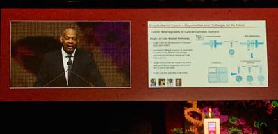
Dr. Carpten closed the session with a talk about the opportunities/challenges for the future of elucidating the complexities of cancer. He focused on the challenges associate!
d with cancer heterogeneity at the population and tumor levels. Noting that < 10% of the TCGA data was from underrepresented populations, he sees a big opportunity to understand the population heterogeneity of cancer, as it relates to environmental and socioeconomic conditions and their effects on epigenetic and transcriptional changes. He then switched gears to discuss intratumoral heterogeneity and the power of single cell genomic analysis to provide a truer picture of heterogeneity compared to bulk methods. He showed data using the Chromium Single Cell CNV Solution in which his team was able to identify the CNV status of just 51 tumor cells in a mix of over 800 normal cells. And, they were able to create individual cell copy number profiles for comparison.
The morning poster session was next on my agenda, and there was a ton of great research to see. "Chromosome-scale haplotyping enables comprehensive discovery of cancer rearrangements and germline-related susceptibility mutations" by Stephanie Greer, Stanford University, Ji Research Group, was the first one on my list. After reading the fascinating blog post she wrote for us last year, "Deconvoluting complex genomic structural variations in metastatic tumors," I had to know more about her cancer study with the Ji Research Group. Using Linked-Reads analysis to study cancer genomes, they were able to access long-range genomic information, making it possible to reconstruct haplotypes and large-scale structural variants. Using this technique, they looked at cholangiocarcinoma, ultimately identifying potential candidate driver genes for that type of cancer.
The next poster, "Structure and evolution of double minutes in a pediatric high-grade glioma," by Ke Xu, St. Jude’s Children’s Research Hospital, focused on brain tumors, specifically the extrachromosomal DNA molecules, called double minutes (DMs), that are often found in them. Using a novel graph-based method, they developed an evolutionary model of DM trajectory. They then applied this model to a pediatric high-grade glioma patient’s disease progression and validated their results using Linked-Reads analysis

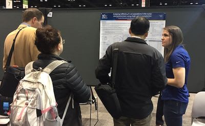
Last up in my morning poster line-up, I took a look at 10x-pert Sarah Taylor’s "Analyzing infiltrating B cell populations in the tumor microenvironment using single cell transcriptomics." It showcased some of the work 10x has been doing with the Chromium Single Cell Immune Profiling Solution, studying tumor-infiltrating lymphocytes to better understand the adaptive immune response. More information about this data can be found in our tumor microenvironment application note.
After that, it was time for the big event of the day, the 10x Exhibitor Spotlight with Miltentyi Biotec, "Single Cell Genomics: Addressing Challenges in Cancer Research." The workshop included two talks about the benefits of single cell analysis in cancer studies, "Delineating Early Tumorigenesis in the Human Breast using Single Cell Genomics" by Dr. Kai Kessenbrock, University of California, Irvine, and "Methods for Sample Preparation and Single Cell Analysis of Solid Tumors" Dr. Jill Herschleb, 10x Genomics.
Dr. Kessenbrock opened the workshop discussing his work using single cell gene expression to delineate cell types and states in the breast epithelium. Using the 10x Chromium Single Cell Gene Expression Solution, the researchers analyzed ~6000 FACS sorted epithelial cells and identified the 3 expected main cell types, each harboring several distinct cell states as seen by clustering using Seurat analysis. A paper describing this work is currently in press and will be out soon. He then focused on his work with the human breast cell atlas (HBCA) team. As part the project, they are evaluating protocols for the preparation of single cell suspensions from breast tissue. During preliminary work, they found 10 of the 11 expected cell types by single cell RNA-seq (the expected adipose cell population was missing). Upon examination of the mechanical dissociation protocol, they realized the adipose cells were lysing and are currently working on a protocol for nuclei isolation for the adipose cells. This highlights the need to optimize sample preparation protocols to minimize batch effects, recover all cell types, and recover high quality live cells as fast as possible.
Following Dr. Kessnbrock, 10x-pert Jill Herschleb presented methods for single cell sample preparation of solid tumors. She started with a workflow and case study for murine tumor dissociation, which included the following workflow: Dissociate with Miltenyi Biotec Ocoto Dissociator → Filter withMiltenyi Biotec MACS SmartStrainer to remove clumps and debris → Lyse RBCs withMiltenyi BiotecRed Blood Cell RBC Lysis Solution → Remove dead and dying cells with theMiltenyi BiotecDead Cell Removal Kit (magnetic beads bind dead or dying cells)
The workflow was tested with a syngeneic mouse tumor model for breast, colon and melanoma (3 replicates each) and the Chromium SIngle Cell Gene Expression solution. Jill and team sequenced 84,000 cells and demonstrated that the workflow and clean- up steps affected library cleanliness (fraction of reads that map confidently back to single cell), library complexity (number of genes expressed/cell), and cell recovery (number of cells seen after sequencing), with increasing number of clean up steps (e.g. RBC lysis and dead cell removal) increasing all of these parameters in the samples tested. Our demonstrated protocol for tumor dissociation for single cell RNA-seq can be downloaded here.
I moved on to the afternoon poster session after the workshop was over. The first poster I looked at was "Sensitive single cell copy number profiling using a novel microfluidic droplet based platform" by Rui Li, McGill University, which described his research using the Chromium Single Cell CNV Solution to sequence and analyze 1 glioma stem cell sample and 2 breast cancer samples. With this method, the genomic basis for tumor heterogeneity could be more fully observed and understood.
My next stop was "Characterization of colorectal liver metastasis at single-cell resolution reveals dynamic interplay in the tumor microenvironment" by Anuja Sathe, Stanford University, where I learned about her use of scRNA-seq to better characterize colorectal cancer (CRC) metastases in the liver. They characterized features of 1,551 cells from 2 CRC liver metastases, which may provide insight into potential therapies for patients.
The final poster for the day was "Single-cell transcriptome analysis on lymphocytes of GATA2 deficiency patients" by Elina Hirvonen, University of Helsinki. In it, Hirvonen describes how they examined the effects of GATA2 deficiency in hematopoiesis. Using the Chromium Single Cell Gene Expression Solution, they observed that GATA2 had an impact on several genes that play a part in immune and cellular mechanisms, which suggests that their research could influence future patient care.
After seeing all the posters, I was looking forward to hearing some talks. I decided to attend two Minisymposia, both of which focused on the use scRNA-seq in developing potential treatments for acute myeloid leukemia (AML). The first was "Single cell RNA sequencing reveals AML immunoediting under pressure from engineered T cell therapy," presented by Kelly Paulson, Fred Hutchinson Cancer Research Center. Dr. Paulson’s team works with targeted T cell therapy, and she discussed the potential of engineered T cells in targeting AML, as well as the challenges. She also described how they are working to overcome difficulties by using scRNA-seq to identify mechanisms that could potentially be targeted to prevent immune escape. Dr. Paulson works with Aude Chapuis, who also gave a great presentation adoptive T cell therapy in our Exhibitor Spotlight on Sunday. Read more about Dr. Chapuis’ presentation here.
Next was "Sequential transcriptomic and phosphorylation landscape of acute myelogenous leukemia (AML) on the single-cell level," presented by Victoria Wang, UCSF. Dr. Wang and her team used a combination of scRNA-seq and mass cytometry (Cy-TOF) to analyze 60 samples from 12 patients during their treatment for AML. They were able to track cellular changes in response to treatment.
Stay tuned for more details about AACR 2018 Day 3.
