Answering your questions about Visium Spatial Gene Expression for FFPE: Spotlight on sample prep
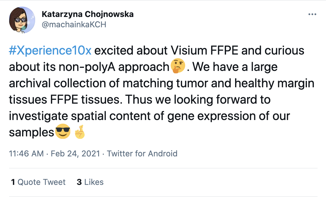
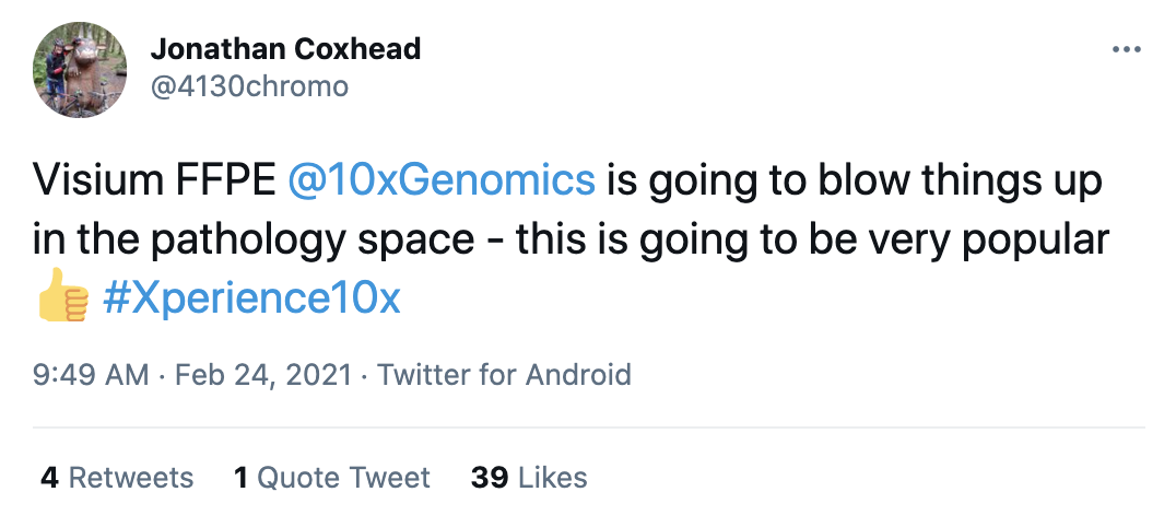
While formalin-fixation, paraffin-embedding (FFPE) is the most common method of tissue preservation, it can damage RNA, making it difficult to perform transcriptomic-level investigations. Back in February, during our Xperience event, when we discussed a new Visium product that would allow users to overcome the limitations of FFPE preservation, we were thrilled to see that many of you were just as excited as us.
Now available, Visium Spatial Gene Expression for FFPE gives you the tools you need to unlock what’s been hiding in your samples. And, with the Visium for FFPE webinar series, we’re making sure you have the skills you need, too. This 3-part series gives you a look into every aspect of our newest assay, from sample prep to data analysis.
Our first session, available to watch on-demand, introduced Visium for FFPE, walking through the workflow and explaining the technology behind it. Using a split-probe approach and RNA-templated ligation to retain high sensitivity and specificity in gene detection, Visium for FFPE combines the benefits of histological techniques with RNA sequencing, letting you spatially profile RNA expression for over 18,000 genes in human and mouse FFPE samples with high resolution across entire tissue sections.
Visium for FFPE is a game changer, enabling comprehensive insights into the spatially resolved transcriptomics of biobanked samples. But to make this analysis possible, there are a few key sample and slide preparation steps that differ from our original fresh frozen assay to keep in mind. We took a closer look at sample prep and workflow considerations in our most recent webinar, Getting Started with 10x Genomics Visium Spatial for FFPE, digging into the important details that will help you get your experiments up and running. 10x-perts Jacob Stern, Senior Product Manager, and Francesca Meschi, PhD, Staff Scientist - Assay Development, took us through the sample prep workflow, sharing their tips and tricks for working with FFPE samples to get the best results, covering everything from RNA quality assessment and imaging techniques to tissue placement and sequencing considerations.
As you might expect, there were plenty of questions for our 10x-perts during the live Q&A session. Keep reading to preview some of the highlights, and then watch the full webinar on-demand.
How it works
What is decrosslinking, and why is it done?
The process to make FFPE blocks includes a formaldehyde fixation, and that causes DNA, RNA, and protein to crosslink in many different ways. In order to make the RNA accessible in the workflow, we need to include the decrosslinking step that makes the RNA molecules accessible again.
Can this technology be used for proteins?
Yes, at this point, it is compatible with immunofluorescence (IF) as well as H&E staining upstream of the RNA workflow. So, Visium detects a handful of proteins at the resolution of immunofluorescence. We're also working on a highly multiplexed protein assay that will use a similar technique to CITE-seq on single cell gene expression. That is in development, and it will allow for much higher plex protein detection on the same tissue section as RNA and morphology.
So from my understanding, you're able to detect 18,000 human genes. Is this 18,000 unique genes or are variants of the same gene included in that number? Where can one find a list of 18,000 genes?
Yes, 18,000 unique genes. We've designed, largely, one probe pair per gene, and the probe pairs are designed to capture as many different transcript isoforms as possible for each of those genes. The probe list, along with the sequences targeted by the probes, is available on our support site right now.
What are UMIs and genes per spot? How does that compare to fresh frozen? How do these numbers extrapolate per cell?
The UMIs and genes per spot relates to the transcripts that we sequence and how those are related to the spatial localization on the slide. Every transcript will have a barcode, which is dependent on where on the slide that molecule was captured. So, we always calculate the number of genes that have been detected, and we also calculate the number of transcripts for that specific gene that were calculated. And those are indicators per cell. In a similar way to how single cell gene expression data is handled, we indicate number of genes and UMIs per cell.
In terms of numbers per cell, that is very tissue-dependent. If you have a tissue where the cell size is very small and so they're all crammed together, you may have anywhere between 5 and 10 cells per spot. If you have a tissue where the cells are bigger in size, you may actually have just one cell per spot. So, again, that is very, very tissue-dependent.
In terms of comparison with fresh frozen, we have seen high correlation. In FFPE tissues with high-quality RNA, we get a sensitivity that is comparable to fresh frozen. Also, in terms of the type of genes that are detected, I do want to caution here against some of these comparisons. What we have seen is that with human tissues, if you compare fresh frozen and FFPE, even if they came from the same donors, they may look different because it's hard to be exactly in the same region, and so that is something to keep in mind when you make a comparison.
Experimental design
This protocol is optimized for FFPE tissue, but you mentioned frozen tissue options. Are we able to use frozen sections or is FFPE preferred?
If you have fresh tissue, I would recommend using the fresh frozen workflow. Why risk the quality of your RNA when there's a solution that's available? But the FFPE assay is compatible with FFPE tissues that are already prepared.
How many ROI, regions of interest, per slide, can be analyzed? What is the maximum area per ROI?
So, we don't we don't really talk in terms of ROIs. We normally talk about Capture Areas. Each slide has four Capture Areas. Each one of these Capture Areas is 6.5 x 6.5 mm, and any portion of that Capture Area that is covered by tissue will yield RNA data, so that whole area can be analyzed. Each of the Capture Areas has 5,000 uniquely spatially barcoded spots. Once you have loaded the data in Loupe Browser, you can actually select specific regions and only focus your data analysis on those regions if that’s of interest for your scientific question.
Is it possible to request for specific genes that may not be included in the 18,000?
At this point, no, but it is on the roadmap. So if that's of interest to you, I'd encourage you to write to support@10xgenomics.com or to your local representative, and we're very eager to hear what you'd be interested in detecting.
Sample prep for Visium for FFPE
Can I use the assay on slides that are already sectioned?
Unfortunately not. The sections have to be placed on the Visium slide, and, not just that, they have to be placed within the Capture Area. Anything that has already been sectioned on a different slide would not be compatible at this point.
How old can FFPE blocks be? Is there a time limitation? How old was the oldest sample that was tested?
The tissue that I showed earlier, for example, is five years old (from 2016). Internally, we have tested blocks that are older than that. I believe we’ve gone back as far as 2008. However, I would like to point out that, more than the age of the block, it is the quality of the RNA that is a determinant of success. You can have older blocks that have been preserved very well and still have intact RNA as measured by a good DV200 score. Or you may have blocks that are just a year old and have highly degraded RNA. So I would encourage you to look at the DV200 score for your block more than its age.
Why make an incision on the tissue? How deep should it be?
If your tissue is larger than the size of a Capture Area, you can make small incisions on the tissue to score it so that when it’s sectioned, the sections are the right size for our Capture Areas. If the tissue is too large for the Capture Area or if there is excess wax, it could potentially invade a neighboring Capture Area, so we definitely want to make sure that whatever we put on the slide is the right size. In terms of depth, you're really gliding that razor with almost no pressure. You just want to go, let's say, 10–20 microns—enough to do a couple sections.
Data analysis
Is there any chance for probe-to-probe dimerization, which may cause the increase of background signals?
With Space Ranger, we will be able to detect off target pairing of the wrong left-hand side with the wrong right-hand side, and those will be flagged as part of the pipeline. For questions like this, I would definitely encourage you to tune in to the July webinar about data analysis for more detail.
Is there a way to get deeper transcriptome data from non-spatial single cell transcriptomics, from neighboring sections?
Yes, for sure. We've seen this technique done quite extensively with the Visium for fresh frozen assay in terms of pairing single cell data with Visium data. I've helped deconvolute the spots, and that technique is applicable for the FFPE solution as well. There are a number of tools that different people in the community have put out, and there's a lot of exciting work being done there.
Register for our final Visium for FFPE webinar, focusing on data analysis, now →