Introducing Visium Gateway: your entry to the world of spatial gene expression

Spatial gene expression profiling allows you to see your tissues in a whole new way with a comprehensive view of the spatial organization of newly discovered cell types, states, and biomarkers. Read this blog post to learn about the power of Visium Spatial Gene Expression and our new Visium Gateway slides.
A picture is worth a thousand words. Imagine where biology would be if microscopes had not existed to allow Robert Hooke to discover cellular structures in cork (1). Where would medicine be without Rudolph Virchow’s observations that cellular changes are at the root of all disease (2), giving birth to the field of cellular pathology? Undoubtedly, the ability to view cells and the tissues they make up with microscopes revolutionized science and medicine.
Traditional histological methods, including hematoxylin and eosin (H&E) staining, are invaluable for examining cellular morphology and how it changes in response to disease. However, these stained tissues still hold many secrets that can help give you a more comprehensive view of your sample’s complex biology. Visium Spatial, powered by spatial transcriptomics technology, allows researchers to access those additional dimensions of information by combining standard sectioning and staining techniques with gene expression and protein co-detection.
Visium Spatial: a wealth of data in every slide
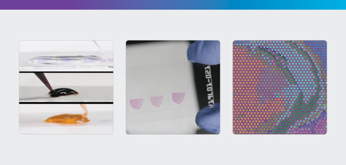
To capture gene expression information from tissue sections, Visium Spatial Gene Expression uses spatially barcoded mRNA-binding oligonucleotides grouped in spots within capture areas on specialized tissue slides. When mRNA is released from processed tissue sections, it binds to capture oligos in the vicinity. A cDNA library that incorporates these spatial barcodes and preserves spatial information can then be prepared from this mRNA. This gene expression data is subsequently layered over a high-resolution microscope image of the tissue section, making it possible to visualize what genes are expressed and where throughout the tissue sample.
With Visium Spatial Gene Expression, researchers have the ability to:
- Perform unbiased, whole transcriptome analysis in morphological context to discover novel insights into normal and diseased tissue
- Combine the crucial insights provided by spatial transcriptomics with the ease and breadth of targeted panels for focused gene expression
- Visualize spatial patterns of gene expression together with protein detection by immunofluorescence on the same tissue section to gain a new perspective on tissue complexity
- Bridge digital and molecular pathology with in situ gene expression to visualize biological signatures, identify predictive biomarkers, and differentiate between normal and diseased tissue in regions of interest
The power of Visium Spatial in action
Profiling gene expression in the context of a tissue sample allows a deeper understanding of biology and disease progression. From tumor microenvironments to brain tissue and beyond, several studies have leveraged the power of Visium Spatial to examine the cellular composition and spatial organization of their samples.
Ji et al. from Stanford University School of Medicine used Visium Spatial Gene Expression as part of a multimodal strategy to compare human cutaneous squamous cell carcinoma (cSCCs) tumors and matched normal skin (3). Spatial analysis revealed the leading edges of these tumors were enriched for basal tumor cells and cSCC tumor-specific keratinocytes, and that TSK-adjacent cells represent a fibrovascular niche. Furthermore, profiling the spatial expression patterns of immune cells in the tumor microenvironment revealed several cell types that likely contribute cSCC immunosuppression mechanisms.
A recent study by Boyd et al. from St. Jude’s Children’s Hospital demonstrated that fibroblast activation is incredibly dynamic in response to viral respiratory infections, resulting in several distinct states—extracellular matrix (ECM)-synthesizing, damage-responsive, and interferon-responsive states (4). Visium Spatial Gene Expression was used to profile lung sections collected from mice 10 days post-infection and understand how these fibroblast populations are organized. The researchers found that damage-responsive fibroblasts co-localized to interstitial inflammation regions at this critical stage of infection response.
In both studies, spatial transcriptomics helped identify the regions where the newly identified cell types reside and function. Additionally, by understanding where these cells are located, the researchers were able to understand how the cells communicate with other cell types in their microenvironment.
Take Visium Spatial for a test drive
As the studies above demonstrate, Visium Spatial Gene Expression provides a comprehensive view of the spatial organization of newly discovered cell types, states, and biomarkers. However, committing to new technology is an important decision, especially when you’re working with a budget. That’s why we’re excited to introduce Visium Gateway slides. Now, you can experience the value of spatial transcriptomics on two tissue samples without stretching your budget.
Explore how Visium Spatial fits into your research with Gateway slides →
References
- L Poppick. Let Us Now Praise the Invention of the Microscope. March 30, 2017. Accessed November 12, 2020. https://www.smithsonianmag.com/science-nature/what-we-owe-to-the-invention-microscope-180962725/
- M Schultz. Photo Quiz. Emerg Infect Dis. 14(9):1480–1481 (2008).
- AL Ji et al. Multimodal Analysis of Composition and Spatial Architecture in Human Squamous Cell Carcinoma. Cell. 182, 1–18 (2020).
- DF Boyd. Exuberant fibroblast activity compromises lung function via ADAMTS4. Nature. (2020). https://doi.org/10.1038/s41586-020-2877-5