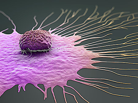
State of the field: Key publications describe how single cell and spatial tools are advancing breast cancer research

From single cell gene expression studies that are beginning to tease out the tumor microenvironment, to atlases of tumor cell types based on localized spatial gene expression—the field of breast cancer research has advanced significantly with the arrival of single cell and spatial technologies. In this post, we acknowledge National Breast Cancer Awareness Month in October by featuring four publications that have used single cell tools to not only learn more about the mechanisms behind the development and metastasis of different types of breast cancer, but also to give us hope as these studies move translational research and clinical trials toward personalized treatments.
The statistics are scary: approximately one out of eight women will develop invasive breast cancer over the course of her lifetime in the US—that’s about 13% of women. Men, too, will be affected: approximately one in 833 will be diagnosed in his lifetime. And, according to the American Cancer Society, the most significant risk factors for breast cancer are being female and getting older—even less comforting, considering we can’t do much about either of these things. Thus, while prevention is key, understanding as much as we can about disease progression and treatment response is paramount.
There are five molecular subtypes of breast cancer, based on the genes that the tumor cells express: hormone-receptor-positive, HER2-positive, and triple-negative breast cancer (TNBC) (1). However, scientists know very little, relatively speaking, about the complex tumor microenvironment (TME) of breast cancers, how tumors interact with the immune system and other surrounding cells and tissues, and what this activity means for prognosis and treatment. As single cell and spatial technologies come into their own, scientists are closing this knowledge gap. Here, we feature four publications in which researchers have made significant advances into defining and characterizing breast cancers on a single cell level.
Creating an atlas: Mapping human breast cancers at single cell, spatial resolution
In the first study (2), “A single-cell and spatially resolved atlas of human breast cancers,” researchers led by Sunny Wu, PhD, of the Garvan Institute of Medical Research, used Chromium Single Cell Gene Expression and Visium Spatial Gene Expression to create a single cell transcriptomic atlas of breast cancer with spatial resolution. Performing single cell RNA-sequencing (scRNA-seq) on 26 primary tumors, they identified all major cell types across all tumors and subtypes. Combining a scRNA-seq–compatible method for intrinsic molecular subtyping, which they developed, with gene signatures identified from their scRNA-seq data, they were able to both classify tumors and show that recurring gene modules (GMs), or sets of genes that are expressed at the same time, lead to the cellular heterogeneity commonly seen within tumors.
Using Visium Spatial Gene Expression on six samples, they saw enrichment of GM3 (EMT, IFN, MHC) and GM4 (proliferation markers) across TNBC samples, while GM1 and GM5 (ER, luminal) were enriched for across estrogen-receptor-positive (ER+) cases. Interestingly, they discovered that GM3 and GM4 phenotypes occur only in regions of breast cancers where the other does not occur. By further describing breast tumors at both single cell and spatial levels, their atlas can be used to improve classification of breast cancers.
Characterizing breast cancer metastases by tracking clonal expansion
In a second publication (3), “The site of breast cancer metastases dictates their clonal composition and reversible transcriptomic profile,” scientists led by Jean Berthelet, PhD, of the Olivia Newton-John Cancer Research Institute, employed an optical barcoding technique alongside single cell gene expression profiling to study the interactive network of clonal cells during the process of metastasis of TNBC, which is associated with worse prognosis and a higher risk of recurrence compared to other breast cancer subtypes.
Performing scRNA-seq on a TNBC breast cancer cell line and the barcoded subclones at two sites of metastasis, lung and liver, they found that metastases were highly polyclonal in lungs but not in liver. Further, subclone transcriptome variability showed that subclones that are dominant in primary tumors remain dominant in the tissue where metastasis occurs. Comparing the gene expression profiles of different barcoded subclones, they identified a metastatic site–driven signature of 1,366 differentially expressed genes; among these, the tumor necrosis factor–alpha pathway was strongly up-regulated in lung compared to liver metastasis. These results suggest that the TME drives the heterogeneity seen in metastases.
Predicting T-cell biomarkers for effective immune checkpoint blockade immunotherapy
In a third publication (4), “A single-cell map of intratumoral changes during anti-PD1 treatment of patients with breast cancer,” Ayse Bassez, a PhD student at the VIB-KU Leuven Center, led scientists to build a single cell map of changes within a tumor using transcriptome and immune profiling. Immune checkpoint blockade (ICB) plus chemotherapy improves treatment response (5), but not all patients respond to neoadjuvant ICB. To explore why, they treated 40 patients with anti-PD1 (a common ICB antibody therapy) before chemotherapy and followed changes within tumors.
Among two cohorts—one that received anti-PD1 and one that received chemotherapy followed by anti-PD1—they performed scRNA-seq and T-cell receptor mapping on matched pre- and post-treatment biopsies using Chromium Single Cell Immune Profiling. Nine patients had T-cell clonotype expansion post-treatment. Using scRNA-seq data while selecting for T-cell expansion and treatment stage, they found that T cells expressing PD1 increase after anti-PD1 treatment. They identified differentially expressed genes between T cells that expand versus T cells that do not expand pre-treatment, discovering that expression signatures in T cells can be used to predict T-cell expansion. Their map of the pre-treatment immune TME that is associated with T-cell expansion after anti-PD1 therapy could offer insight into clinical biomarkers.
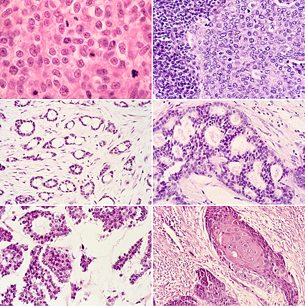
Resolving heterogeneity in DCIS, an early stage of breast cancer
In a fourth publication (6), “Genomic profiling reveals heterogeneous populations of ductal carcinoma in situ of the breast,” Satoi Nagasawa, MD, PhD, at the University of Tokyo, led a team of scientists who used Visium Spatial Gene Expression to reveal cellular diversity within breast cancer tumors. Ductal carcinoma in situ (DCIS) is an early stage of breast cancer that may develop into invasive ductal carcinoma. However, it is difficult to determine who is high-risk and requires surgery, making the determination of risk factors essential to assessment and treatment.
In this study, they first selected and validated genomic risk factors for DCIS using traditional methods, landing on GATA3 and PIK3CA mutations. By performing spatial transcriptome analysis of DCIS using three specific cases, they showed that DCIS cells with GATA3 mutations can sometimes progress to invasive cancer. However, DCIS cells with PIK3CA mutations did not lead to cancer. In conclusion, more accurate classification of DCIS using new markers is crucial for deciding on the right treatment.
Moving toward improved diagnosis and treatment
It has become clear that single cell and spatial research tools are advancing our understanding of breast cancer development, progression, and treatment beyond what we could have dreamed of using traditional methods. Whether making use of newly created spatial transcriptome maps or using gene expression and immune profiling to better describe disease state, clinical researchers are increasingly armed with methods to better diagnose, stratify, and offer individualized therapies for breast cancers.
References:
- https://www.breastcancer.org/symptoms/types/molecular-subtypes
- Wu SZ, et al. A single-cell and spatially resolved atlas of human breast cancers. Nat Genet 53: 1334–1347 (2021). doi: 10.1038/s41588-021-00911-1
- Berthelet J, et al. The site of breast cancer metastases dictates their clonal composition and reversible transcriptomic profile. Sci Adv 7: eabf4408 (2021). doi: 10.1126/sciadv.abf4408
- Bassez A, et al. A single-cell map of intratumoral changes during anti-PD1 treatment of patients with breast cancer. Nat Med 27: 820–832 (2021). doi: 10.1038/s41591-021-01323-8
- Schmid P, et al. Atezolizumab plus nab-paclitaxel as first-line treatment for unresectable, locally advanced or metastatic triple-negative breast cancer (IMpassion130): updated efficacy results from a randomised, double-blind, placebo-controlled, phase 3 trial. Lancet Oncol 21: 44–59 (2020). doi: 10.1016/S1470-2045(19)30689-8
- Nagasawa S, et al. Genomic profiling reveals heterogeneous populations of ductal carcinoma in situ of the breast. Commun Biol 4: 438 (2021). doi: 10.1038/s42003-021-01959-9