Atera In Situ is uncompromised spatial.
An abridged history of spatial biology
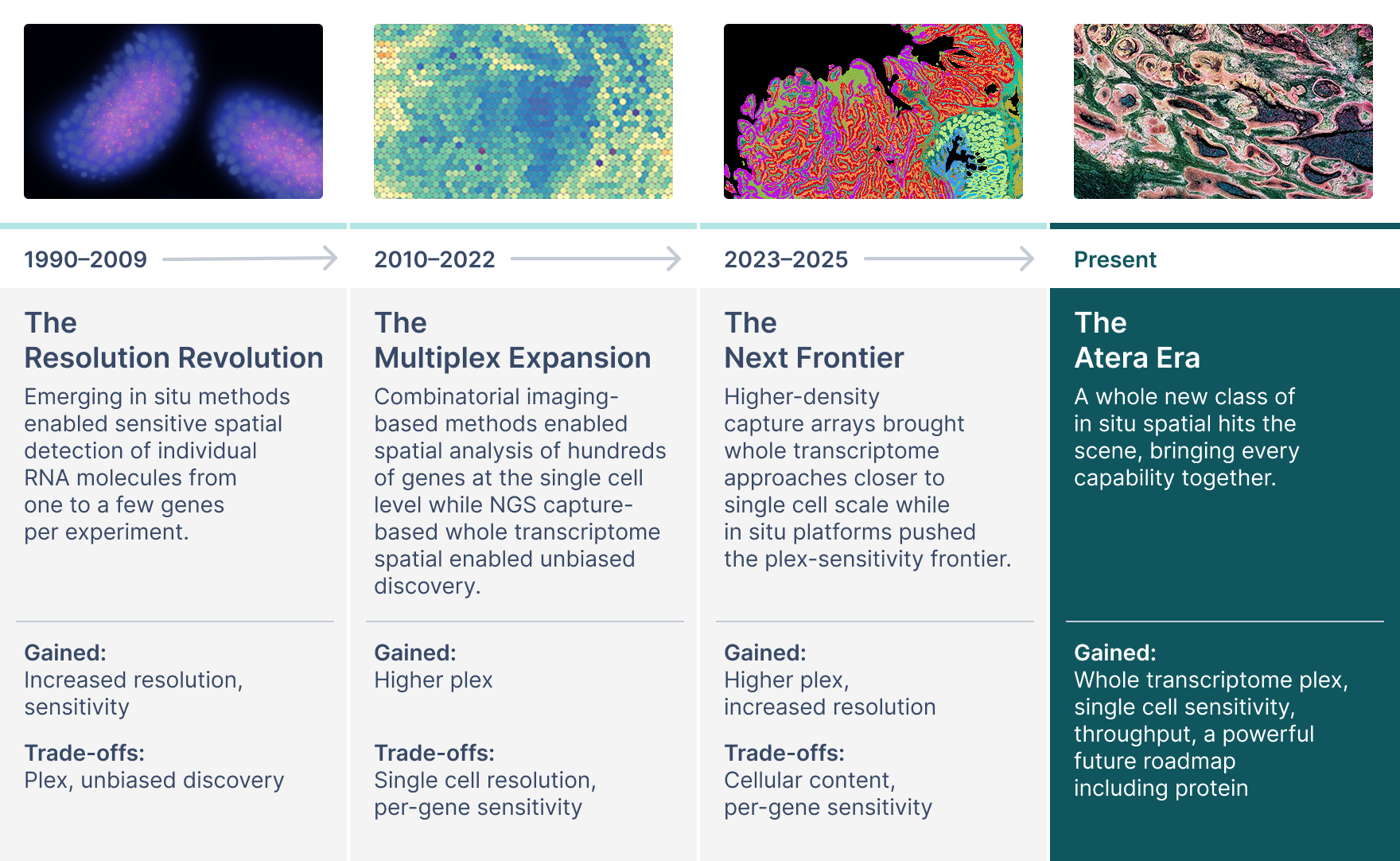
Five years ago, tradeoffs in spatial transcriptomics technology were the accepted norm. You chose the technical capability that mattered most to you, because no platform could do it all.
For example, on NGS platforms you could perform targeted spatial gene expression analysis within a region of interest (ROI), trading an incomplete view of your tissue section and the transcriptome for a sensitive readout of your genes of interest. Or, you could perform spatial whole transcriptome analysis on your whole tissue section for a more complete view of the biology of the sample, but at the cost of lower gene sensitivity. The same compromises played out as spatial imaging technologies developed—lower plex in situ options offered highly sensitive, specific transcript detection, but at the cost of not seeing it all.
Tradeoffs on all fronts were normal: in spatial resolution, plex, panel customization, tissue throughput, assay sensitivity and specificity, speed of runs (if instrument-enabled), and data access. At each evolution, the platforms advanced capabilities, but spatial transcriptomics was still incomplete (Figure 1).
What is Atera?
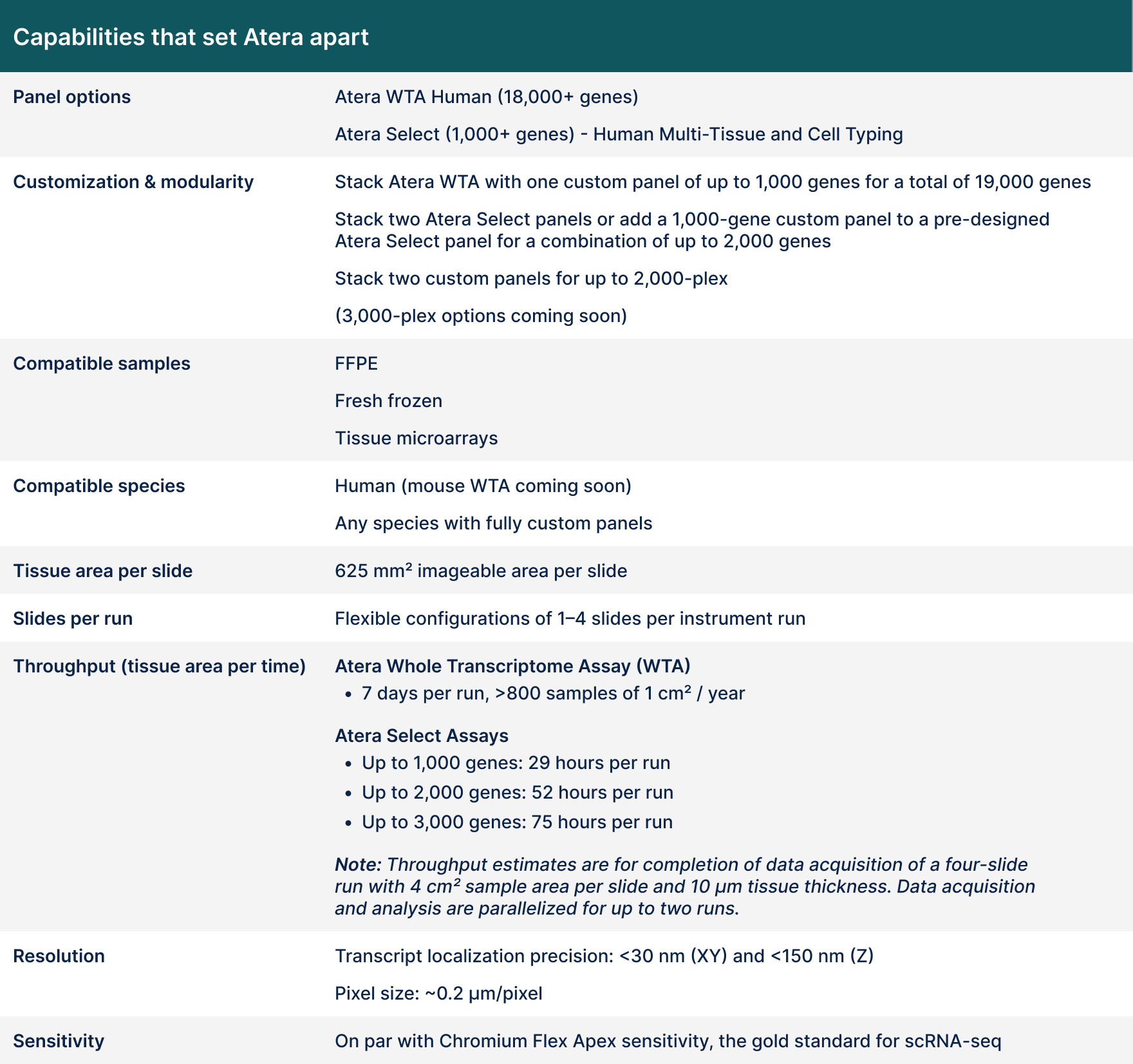
It’s whole transcriptome plex, with customizable and pre-designed mid-plex panels.
It’s subcellular resolution with the sensitivity to match single cell sequencing—think thousands of transcripts per cell.
It’s flexible throughput that also lets you max out your experiments (2,500 mm² of tissue per week, 800 whole transcriptome 1 cm2 samples/year).
It’s interpretable data the moment your run is finished.
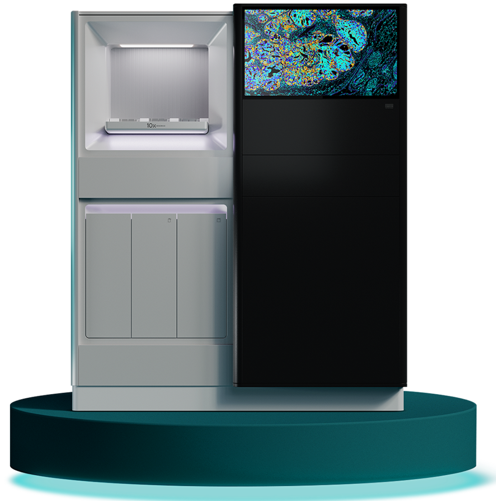
Atera (Figure 2) offers throughput, WT plex, and sensitivity all on the same platform (Table 1).

See inside the Atera platform
Take a deep dive with this interactive demo of Atera and the assay components.
How does Atera work?
Built upon the pioneering Xenium technology, Atera provides one universal workflow (Figure 3) for whole transcriptome spatial insights from fresh frozen or FFPE tissue.
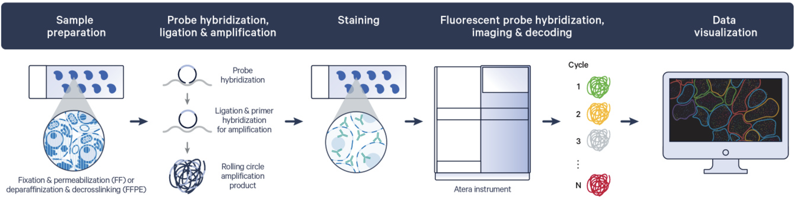
First, you section tissue on a glass slide. Then run the assay with your gene panel(s) of choice. Atera leverages a probe-based rolling circle amplification chemistry, so for the genes that are part of your panel (whether that’s 18,000+ or 1,000+), highly sensitive, specific probes will bind to transcripts in your tissues, ligate, and then amplify the transcript product on-slide using a universal workflow. After cell segmentation staining, you load slides onto the instrument where fluorescence imaging, decoding, and analysis is performed simultaneously. What you get after run completion: a ready-to-explore, spatially resolved, gene expression map at subcellular resolution.
Go deeper into the Atera workflow with these resources:
- Atera Gene Expression User Guide
- Atera Instrument User Guide
- Atera Tested Tissues
- Demonstrated Protocol for Fresh Frozen Tissue Prep
- Demonstrated Protocol for FFPE Tissue Prep
- Demonstrated Protocol for Post-Atera H&E Staining
Sentira Spatial Analysis launches with Atera
Sentira Spatial Analysis is a cloud-based spatial analysis software solution built in a 10x web browser. After on-instrument primary and secondary analyses, data is egressed into Sentira, where you can run compute-heavy pipelines in minutes (compared to hours), visualize and explore data in real time, and share datasets through a simple project invite—all without having to build the computing infrastructure needed yourself.
Learn more about the vision behind Sentira with our press release, and find out how to join the Early Access program.
Your peers answer: What can I do with Atera that I couldn’t before?
“Atera enables us to generate data across larger cohorts at a pace and depth that was previously not practical.” — Jan-Philipp Mallm, PhD, German Cancer Research Center (DKFZ)
[Atera] is able to balance whole transcriptome applications while still maintaining high sensitivity. Take these advances and combine them with the system’s increased throughput—the potential for large cohort studies is incredible.” — Hongkui Zeng, PhD, Allen Institute
"We expect Atera to fundamentally expand the scope of our translational research and accelerate target discovery in inflammatory and fibrotic diseases." — Kevin Wei, MD, PhD, Brigham and Women's Hospital
Atera makes the impossible, possible. Scaling to your biggest biobank studies, defining an elusive disease mechanism or pathway of therapeutic resistance, finally validating a target that no spatial platform could trace definitively, and making the most of each precious sample.
Let’s take the tumor microenvironment as an example. In the past, limited panel size or transcript sensitivity may have blocked your ability to derive clinically relevant insights, like what are the best potential indicators of tumor progression or therapeutic response. But greater biological recovery with a whole transcriptome assay and thousands of transcripts per cell can reveal:
- Which drugs affect the tumor microenvironment in this tissue niche?
- What mutations are associated with disease heterogeneity?
- What immune response biomarkers are near tumors?
Imagine advancing your initial findings to a larger cohort for further validation. Atera throughput could help you achieve the necessary data scale to make actionable conclusions:
- Based on pathway changes to this model system, which drug targets should we move forward?
- How do cell states on the immune invasive front change after dosage?
- Does my antibody localize to the antigen I expect? Are there off-target effects?
What if you wanted to find biomarkers for neurodegenerative disease or better understand the underlying mechanisms of therapeutic resistance in Alzheimer’s? With meaningful biology from large cohorts of clinical FFPE samples and the reliability of a consistent, automated prep, the following insights are within reach:
- What biomarkers in this cohort indicate a treatment response?
- Will this target pass clinical trials?
Atera provides capabilities that maximize your samples and answer your hardest questions.

Tracing clonal evolution and immune response in the tumor microenvironment
Learn how a Stanford research group used Atera to infer copy-number alterations at single cell resolution to reconstruct tumor clonal evolution in breast cancer, and how they followed the changing immune response in situ.
Getting started with Atera
How can I access Atera?
If you can’t get your own Atera system now, you don’t have to. There are other options to access Atera:
- Catalyst Research Services. Submit samples directly to us, including projects as small as a single slide. We process your samples and within four weeks of project start, you'll get Atera data back. 10x bioinformatics experts are also available.
- Instrument leasing and reagent rental programs. Speak to an Atera sales specialist about our leasing options, which reduce upfront capital requirements while maintaining instrument access.
Where can I find Atera support resources?
Find support resources for Atera In Situ here. Other helpful resources to get started include:
Product specs and overview
Experimental design and workflow
- Atera Experiment Builder
- Panel Design Guide
- FAQs (Technical)
Data analysis resources

Take the next step with Atera In Situ
Talk with a technology specialist about Atera In Situ to get started.