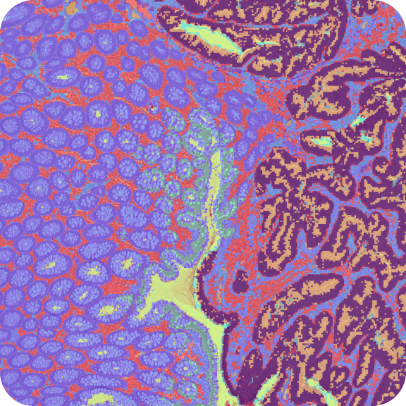
Visium HD 3' Gene Expression Library, Human Ovarian Cancer (Fresh Frozen)
HD 3’ Spatial Gene Expression dataset analyzed using Space Ranger 4.0.1

Learn about Visium Analysis
Biomaterials
Human ovarian cancer tissue was obtained from BioIVT.
- Sex: female
- Age: 39 years
- RIN 9.91
Sample preparation
A 10 µm section was taken with a cryostat (Epredia CryoStar NX70). Tissue preparation, sectioning, H&E staining, and imaging followed the Visium HD 3' Fresh Frozen Tissue Preparation Handbook (CG000804).
Imaging
- Image type: H&E
- Microscope: Olympus VS200 Slide Scanner
- Objective magnification: 20X
- Numerical Aperture: 0.8
- ScopeLED light source: VS200 LED, Olympus integrated bright field source
- Camera: iDS VS-264C, Olympus scanner integrated camera
- Exposure: 500 microseconds
Assay workflow
Library preparation followed the Visium HD 3' Spatial Gene Expression User Guide (CG000805).
- Slide serial number: H1-N3KXRGQ
- Area: A1
- Instrument: Visium CytAssist
Sequencing
- Indexing: Dual index plate TT set A; sample index G6
- Sequencing instrument: Illumina NovaSeq 6000
- Sequencing configuration: 43 bp read 1, 75 bp read 2, 10 bp i7 sample index, and 10 bp i5 sample index
- Sequencing depth: 558.7 million read pairs
Analysis
Space Ranger v4.0.1 was used to map FASTQ files to the reference, detect tissue, align the data to the microscope and CytAssist images, segment cells, and output feature-barcode matrices for further analysis.
How to view data
To get started, download Loupe Browser v9.0 or later to explore the Loupe file, or read more about the other Space Ranger outputs.
This dataset is licensed under the Creative Commons Attribution 4.0 International (CC BY 4.0) license. 10x citation guidelines available here.