GEM-X Epi Multiome (previously known as Chromium Single Cell ATAC + Gene Expression) provides a dual readout of chromatin accessibility and gene expression from the same nuclei, revealing how epigenetic regulation affects cellular identity and function. This blog introduces the benefits of this experimental approach, a basic overview of how the Epi Multiome assay works, experimental and cost efficiencies enabled by GEM-X technology, and how you can get started.
Searching for the “why” behind cell state and function
You have a nagging question. You can already describe distinct cell states from your transcriptome data, but you're missing the why. That’s something the transcriptome alone isn’t able to answer for you.
- Why do metastases from the same primary tumor activate completely different gene programs and respond differently to therapy?
- Why are certain neuronal and glial populations selectively vulnerable to neurodegenerative disease while others are spared?
- Why does T-cell immunity, such as responsiveness to pathogens or differentiation potential, wane with aging?
These questions can’t be fully answered without understanding the layer of biology behind gene expression.
Epigenetic regulation—it’s the software that controls the hardware of DNA; a program that dictates which specific genes to express or mute, rewriting its instructions in response to environmental signals, cellular interactions, or intracellular cues (Figure 1).
It looks like DNA methylation sites that block transcriptional machinery. Or histone proteins that keep DNA tightly bound, preventing access to certain regions for expression. These epigenetic features, among others, provide a dynamic rule book that controls gene expression and thus individual cell state and function.

If you have a nagging question about cellular identity—why is a certain cell state in your biological system acting the way it is—could a view of epigenetic regulation fill that gap?
Why same-cell multiomics
You want to know more about your cells and samples. Intuitively, you understand the value of multiomics, seeing multiple biological layers at the same time. But you also want the best information possible, so you can build hypotheses and conclusions on accurate data. That’s why direct linkage of multiomic readouts is preferable to inferred linkage.
For example, you could infer the relationship between epigenetic regulation and gene expression. You could run separate single cell RNA-seq and ATAC-seq assays, then computationally integrate the two datasets. But this could introduce some errors:
- Mismatched cell types across modalities, especially rare or transitional states
- Misattributed chromatin–expression links (accessibility doesn't always predict expression, and can precede it)
- Loss of single cell resolution, since data gets averaged to cluster-level for comparison
In a GEM-X Epi Multiome experiment, you get both the chromatin accessibility profile and the transcriptome from the same nucleus. This means you can directly ask: "In this specific cell, is this peak open, and is the gene nearby actually being expressed?" The correlation is directly observed—it’s ground truth at the single cell level.
How do you measure chromatin accessibility and gene expression from the same nuclei?
To get you started, we wanted to break down the basic steps of the GEM-X Epi Multiome workflow (Figure 2) and point out some tools that can help you succeed.
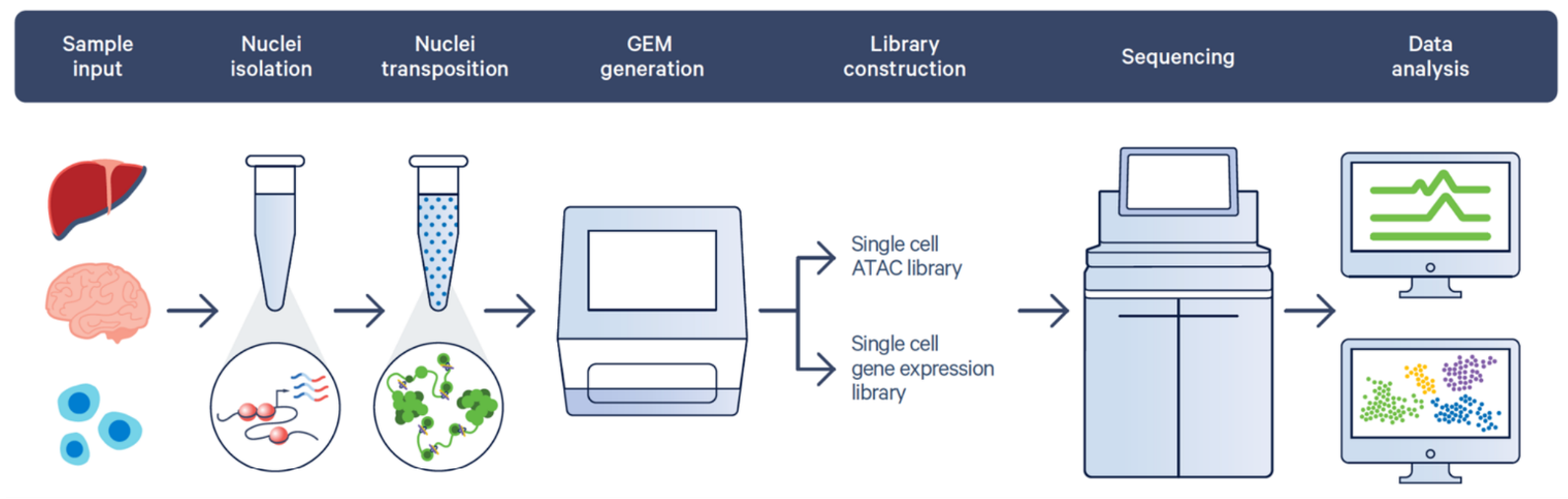
First, sample prep. You can start with cells or tissue, but, at the end of the day, you need nuclei because that’s where the chromatin accessibility measurement will come from. Nuclei isolation can be tricky, but we’re optimizing that with our Nuclei Isolation Kit, which allows you to generate a nuclei suspension from a wide range of frozen tissue, little to no optimization required, in ~1 hour.
Using the Nuclei Isolation Kit is usually the most straightforward course for your sample prep, though certain tissues may need a different approach. Check out our tested tissue list:
- Nuclei Isolation Kit Tested Tissues
- Nuclei Isolation Kit Sample Prep User Guide (for when you’re ready to get started!)
If the Nuclei Isolation Kit isn’t the right fit, we have extensive user guides and protocols to help you optimize for different sample types:
Following nuclei isolation, you can take steps to generate a single cell ATAC library and a single cell gene expression library. You first incubate your nuclei with transposase, which enters the nuclei and fragments the DNA in open regions of chromatin (revealing the places primed for gene expression). In subsequent steps, those transposed nuclei get individually partitioned with Gel Beads that can barcode both the DNA fragments and mRNA from each nucleus. The GEM (Gel-Bead-in-emulsion) includes reverse transcription reagents to generate cDNA. After incubating the GEMs, you get barcoded DNA and barcoded, full-length cDNA to generate ATAC and gene expression libraries from the same nucleus.
See the full process broken down step-by-step in our assay user guide, including a web version:
- GEM-X Epi Multiome User Guide
Analyzing your GEM-X Epi Multiome data
After library prep and sequencing, it’s time to analyze your data and glean insights from a direct, same-cell measurement of chromatin accessibility and gene expression. The Cell Ranger Arc pipeline has you covered to process raw sequencing data, then Loupe Browser software does the visualization. These tools enable detailed analysis of gene expression, chromatin accessibility, and their interconnections.
But what are those connections, and what do they look like? These Loupe Browser tutorials help you get a feel for what Epi Multiome data looks like in the tool’s interface, providing an overview of multiome data concepts (like gene–peak feature linkages) with visuals and walk-through instructions.
Take a look at our Analysis Guides for Epi Multiome data to go deeper into some important concepts:
- Next Steps in Epi Multiome Downstream Data Analysis
- Quality Control and Filtering of 10x Genomics Epi Multiome Data
The 10x difference
GEM-X Epi Multiome has a couple important differentiators grounded in its improved microfluidic technology and assay chemistry:
- High capture efficiency: Profile up to 80% of the nuclei from your original nuclei suspension to make the most of a hard-earned sample prep and maximize insights from precious clinical samples.
- Lower costs: Throughput improvements pair with a two-fold increase in gene expression sensitivity over previous chemistries to lower costs per cell—because you can run more cells in an experiment and use less of your budget on sequencing.
- More discovery power: Capturing more genes per nucleus also means you can effectively detect lower-abundance transcripts and rare cell populations.
Beyond the assay itself, you get the expertise of the 10x Genomics team and an ecosystem of resources. Our best-in-class support team has >90% satisfaction rate, and we provide extensive documentation and training to guide you through the whole process, from sample prep to data analysis.
More helpful links for getting started
Learn more about GEM-X Epi Multiome on our product page. Explore some of the links below to find other helpful resources:
300+ publications using Epi Multiome assays
Product specs and overview
Experimental questions
- How-To Videos
- FAQs (Technical)
Data analysis resources

Take the next step with GEM-X Epi Multiome
Talk with a technology specialist about GEM-X Epi Multiome to get started.