Predicting disease flares in systemic lupus erythematosus using single cell transcriptomic analysis

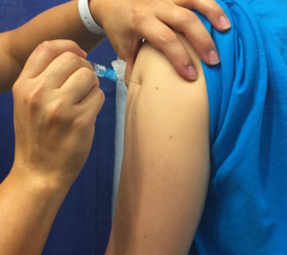
To honor Lupus Awareness Month in May, we feature a research publication that makes use of 10x Genomics Single Cell Gene Expression to study the shared baseline gene expression signatures between vaccine response and the flares that occur with one type of lupus (1). Uncovering the cellular activity that contributes to these flares by comparing the underlying immune response to similar activity after vaccination could point to biomarkers of disease and new therapeutic targets.

Lupus is an autoimmune disease in which a person’s immune system mistakes the body’s healthy cells as foreign and attacks them, causing rashes, pain or swelling of the joints, extreme fatigue, and other symptoms. Similar to other autoimmune diseases, lupus is difficult to diagnose and treat—symptoms can vary among individuals, resemble other diseases, and come and go over time. The most common type of lupus is systemic lupus erythematosus (SLE), which causes pain and swelling possibly all over the body.
Because clinicians want to be able to predict who might develop lupus and how and when flares may occur, focusing on immune response to vaccination—which varies widely across individuals, with some people exhibiting a greater immune response or higher antibody production than others—may offer insight into immune response in autoimmune diseases like lupus. Immune responses, in general, vary among people, and there is growing evidence that the state of the peripheral immune system before vaccination or disease can predict subsequent immune response (2). Interestingly, vaccines, including influenza, yellow fever, and others, and infections both cause an increase in plasmablasts in the blood. Plasmablasts have also been shown to increase with flares in SLE (3). In a recent study led by John Tsang, PhD, Co-Director, NIH Center for Human Immunology (CHI), he and his team built on previous work by using SLE as a model to investigate common baseline gene expression signatures associated with vaccine response in healthy people and disease activity in SLE.
Predicting vaccine response and lupus flares with the same baseline signature
In previous work, Dr. Tsang and team pinpointed what they called a baseline (before vaccination), blood-based gene expression signature of 10 genes that they found were predictive of antibody response to the influenza vaccine in several but not all of the studied clinical trial cohorts (4). When they applied this 10-gene signature (TGSig) to peripheral blood mononuclear cell (PBMC) expression data from another clinical trial of the yellow fever vaccine, which is different from the inactivated seasonal influenza vaccine in that it uses a live attenuated virus, he found that TGSig could also predict antibody response to yellow fever vaccination. When he applied TGSig to a pediatric SLE cohort, he found that TGSig could also be tied to baseline disease activity in SLE and that flares (characterized by an elevation in plasmablasts) could be predicted based on the expression of these 10 genes.
Drilling down to the cellular level: Single cell analysis reveals activated network
In a previous NIH cohort study, the predictive 10 genes correlated with the frequency of one specific subpopulation of B cells—those that expressed CD20 and high levels of CD38. In order to determine exactly which cell subpopulations expressed the signature genes, they used multiomic cytometry to simultaneously profile gene expression and 82 cell surface proteins of 53,201 single cells from healthy high and low flu vaccine responders in the NIH cohort. Results revealed that the bulk gene signature reflects the extent of activation in multiple immune cell populations at baseline, including plasmacytoid dendritic cells (pDCs) and lymphocytes, specifically cells involved in a plasmacytoid dendritic cell/type I interferon/T and B lymphocyte network.
The activation of these molecular circuits have already been shown to be involved in the progression of SLE and other autoimmune diseases (5), and this study offers further support that this increased activation during a non-flare state may point to higher plasmablast-associated disease activity. In short, activation of pDCs and B and T cells in both vaccination response and non-flare state of SLE can be used to predict antibody response levels and flares, respectively. Future studies may look at how this signature can be used to predict immune response activity in other autoimmune diseases outside of lupus.
Driving toward biomarkers and therapies for lupus
The study’s findings suggest that immune response and disease response in some autoimmune conditions can be predicted based on a common baseline set of marker genes, and that flares in SLE may involve specific cells that can be targeted as either disease biomarkers or as therapeutic targets. This type of single cell analysis may go a long way toward not only managing symptoms of lupus and other autoimmune conditions, but also developing clinically useful blood-based biomarkers or discovering new pathways for targeted therapies.
Learn more about single cell analysis for immunology by exploring the getting started guide or visiting the 10x Genomics Immunology Gateway.
To connect with a global community of immunologists and discover the latest in immunological research, register for the 10x Genomics Global Immunology Virtual Symposium, held May 24–28.
References:
- Kotliarov Y, et al. Broad immune activation underlies shared set point signatures for vaccine responsiveness in healthy individuals and disease activity in patients with lupus. Nat Med 26(4): 618–629, 2020.
- Tsang JS. Utilizing population variation, vaccination, and systems biology to study human immunology. Trends Immunol 36: 479–493, 2015.
- Banchereau R, et al. Personalized immunomonitoring uncovers molecular networks that stratify lupus patients. Cell 165: 551–565, 2016.
- Tsang JS, et al. Global analyses of human immune variation reveal baseline predictors of postvaccination responses. Cell 157: 499–513, 2014.
- Banchereau J & Pascual V. Type I interferon in systemic lupus erythematosus and other autoimmune diseases. Immunity 25: 383–392, 2006.