Why I chose spatial: The what and where of regrowing limbs
Impact at a glance: The Visium Spatial platform allowed one of the leading experts in regeneration biology, Dr. Elly Tanaka of the Research Institute of Molecular Pathology, to study neuroregeneration in axolotls, including looking at changes in cellular transcriptomes over time next to injured tissues. Her team’s work seeks to understand the molecular basis for why animals respond differently to injury and discover a link between pathways present in regenerative animals that can be implemented in non-regenerative animals.
“And so there, again, the spatial information was very important for us to develop a map of the forebrain of the axolotl and understand the cell types that are there.”
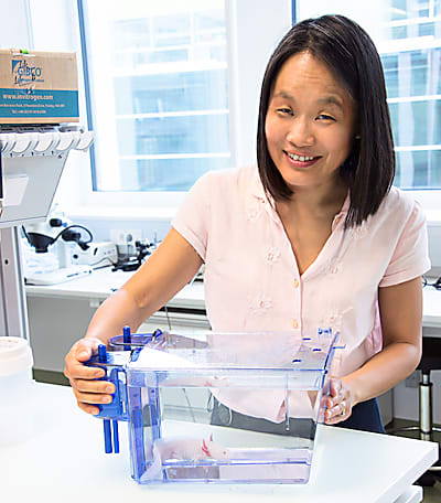
Regeneration—growing back digits, limbs, and even whole organs—can seem like the stuff of science fiction. Unless you happen to study developmental biology in the axolotl, like Elly Tanaka, PhD, Senior Scientist at the Research Institute of Molecular Pathology, who was described as the leading expert in regeneration biology in 2017 when she was awarded the Ernst Schering Prize.
We recently highlighted her neuroregeneration work in the axolotl and, for the latest entry in our Why I Chose Spatial series, we were lucky enough to get a full interview with her. Read on to see how she got into science, what drove her interest in regenerative biology, and how spatial gene expression technologies have shaped her research.

How did you originally get interested in science, and in your specific field?
In high school, I enjoyed both English and sciences. During college, I felt more comfortable with sciences. Eventually, I ended up in biochemistry. What got me interested in doing lab research was that one summer I went out to Seattle, and the only job I could get was in a lab. So I was working with worms, and thinking about sex determination. And that convinced me that it was a good life for me.
In graduate school, in addition to the cytoskeleton work I was doing, I was always reading about developmental biology. And I was very interested in these fascinating experiments where you can cut an embryo in two, and then the two halves will develop into entire embryos.
By the time I was finishing graduate school, I bumped into the regeneration literature in salamanders, and I thought, ‘Wow, what an amazing example of regulation that might have medical implications one day!’, and so then I was hooked.
That's how I decided to go into regeneration research, and [I went] to London to Jeremy Brockes' lab to learn how to work on regeneration.
We did a piece on your earlier work, so would you mind describing your current research projects and what you’re trying to answer in the regeneration space?
In regeneration, we're interested in how removing or injuring a piece of tissue generates a local response in the surrounding cells that causes the cells that are there to undergo proliferation and lose their adult phenotype. They then undergo the process of differentiating again in order to replace the missing tissue.
We're now doing that in a number of tissues. Originally we were working on this in limb and spinal cord regeneration, but we branched out into the brain, which was the first instance where we applied spatial transcriptomics. We are also now looking at heart regeneration. Regeneration is a complex thing to study, because it's an adult tissue that has many, many different cell types. Then you injure it, and there are so many genes that change, and there's an inherently local aspect to it.
We know from classical experiments that the cells that contribute to the regenerating tissue are all cells that come within several hundred microns of the amputation plane. So spatial organization and spatial responses are extremely important in studying regeneration. When we had to grind up tissue and do normal bulk RNA-seq, we were not only integrating all the different cell types and pushing them all into a tube, we were collecting bits of normal tissue together with the responding tissue.
In that way, spatial transcriptomics has been extremely important for us to look transcriptome-wide at what changes are going on, but really specifically in those cells that are next to the amputation plane. And because of single cell sequencing, we have cellular fingerprints of which cell types are expressing what transcriptomes. So, although spatial transcriptomics doesn't necessarily have single cell resolution, we can say that, in this area, there's these two or three cell types. Then, by saying that we have these two or three cell types in this one area, we can hypothesize that these cell types are probably interacting as part of the regeneration response. Then we can look at which signaling pathways are starting regeneration.
One advantage of the axolotl system is that the cells are 10 times the size of human cells. So we get a big advantage in single cell transcriptomics and we’re almost at single cell resolution with the [Visium] platform. So it's been a very nice system for us to be able to look at the wave of changes in cell transcriptomes that are happening in a time course of regeneration next to injured tissue.
One of our next questions was going to be what led you to consider using spatial technology to answer your research questions, but your comment about investigating cells within a few hundred microns of the injury site kind of answered that!
We used it not only for regeneration but in the first paper where we were also characterizing the axolotl forebrain. It really helped us to identify which cell types were in which part of the brain. People have looked at single markers to try to make conclusions about which part of the axolotl brain was homologous to other mammalian brains. But it turns out that single markers are just not good enough.
When you have a fingerprint transcriptome, then you can cross-compare cell types between different animals and make better hypotheses of their homologous cell types. We performed Visium on the axolotl forebrain, with the single nuclei sequencing data that we have, and then we could map those cells with Visium. That could help us confirm whether the cell types that we had seen were homologous to certain cell types in reptiles and mammals, and whether their location in the brain actually makes sense.
So why did you choose Visium? And did you have any unique challenges trying to do these neuroanatomical and spatial analyses in amphibians versus, say, mammalian organisms?
We chose Visium because it’s an undirected technology in the sense that you sequence what’s there, so we didn't have to design a probe set in order to do this work. Our first Visium experiments were done with Barbara Treutlein, PhD (ETH Zurich, Switzerland), who was the one that implemented the technology. Since then, we have used it on other axolotl tissues and it is set up here at the Vienna Biocenter Core facilities.
After these first experiments, based on the characteristics in terms of reliability and sensitivity, it seemed like it was going to be the best option for us, especially because we have so many technical development things going on in our lab that the thought of trying to set it up ourselves from scratch was just way too much. For all those reasons, Visium seemed like the best platform.
As we said, since we [axolotls] have cells that are 10 times larger than mammalian cells, the resolution was fine for us. For technical challenges, actually getting the lysis conditions. I think everyone faces this; I don’t think it’s axolotl specific, but it’s probably tissue specific where you put tissue on the slide and then you have to determine a lysis condition. That took some trials to get data where we were confident we were getting efficient extraction in most regions of the section we were interested in.
What are the most interesting things that you’ve found with Visium?
People are interested in the forebrain of animals, because it's the part that forms our cerebral cortex, which is a big part of higher thinking. There was always a big mystery, whether salamanders had a homologous part or not, whether they had a cortex-like area.
By doing single nuclei sequencing classification of cell types, and then mapping those onto the Visium data, it became clear that a large part of the forebrain of the axolotl, which could have been the cortex, seemed to be like the olfactory cortex area in mammals. But then there's a little corner of the brain that has this one cluster phenotype that would be consistent with being the dorsal cortex, which I found kind of fascinating. Imagine how that might have, over evolution, somehow expanded. It’s just kind of fun to think about.
Where do you plan on taking your research next, and how will spatial analysis keep contributing to that?
On one hand, we’re implementing Visium to understand pathways that might be contributing to heart regeneration in the axolotl that might not be present in other animals. And we're interested to see whether we can somehow make the link between pathways present in a regenerative animal and if we can implement them in non-regenerative animals and get better regeneration.
We are also interested in these probe-based approaches, like Xenium, for some mouse bone regeneration work. It would be very interesting for us to see cohorts of genes being turned on after bone injury and where cells are and what phenotypes they are, at higher resolution. It might be very good to look at equivalent injuries in axolotl, and then again, ask what's present or missing in the axolotl, what is present or missing in the mouse, and then try to really understand the molecular basis for why animals respond differently to injury.
We love to hear scientists talk about their work, so was there anything really interesting—in this paper or otherwise, spatial or not—that you want people to know about?
We've done a lot of single cell sequencing also. One of my favorite experiments was when we compared limb regeneration in axolotl with limb amputation in frog. Frogs can regenerate the limb before metamorphosis, but after metamorphosis they don't regenerate or form a blastema. They grow this kind of cartilage spike, but they don't regenerate it within. And so we used 10x Genomics’ sequencing to do a time course of these two processes.
With the axolotl, we've shown that fibroblasts dedifferentiate into limb bud progenitor-like cells. In essence, they go back in time to become like a limb progenitor. And when we looked at the frog cells and looked at their molecular fingerprints, they didn't look like they were going back, actually, as completely to developmental cells. We didn't know whether that's because there was something inhibiting them from doing it in the environment, or they had some intrinsic inability to reenter that program.
So Tzi-Yang Lin, the graduate student who did that work, he worked out transplanting these frog cells back into a frog limb bud in a most permissive environment. So he transplanted these genetically labeled cells, these adult frog fibroblasts and also frog blastema cells, back into the limb bud. Then we dissociated that limb bud and performed single cell sequencing. And we could distinguish the donor and host cells because of GFP and mCherry expression. And what was beautiful, what was so clear, was that the transplant itself did not change their phenotype. They looked much more like the original phenotype, and didn't seem to adapt almost at all to the embryonic environment and didn't come close to the embryonic cells. For us, this was a very convincing experiment that showed that there's some intrinsic block, probably at the chromatin level, that the frog cells have that keeps them from being able to become like a developmental limb cell again. I think that was a super cool experiment!
This interview has been edited for length and clarity.
To learn more about Dr. Tanaka’s work, please visit her lab’s website; or read more about Visium Spatial Gene Expression here.
Want to find out why other researchers have chosen spatial? Keep reading the series!
