Mixture of Healthy and Cancer FFPE Tissues Dissociated using Miltenyi FFPE Tissue Dissociation Kit, Multiplexed Samples, 4 Probe Barcodes (Next GEM)
Flex dataset analyzed using Cell Ranger 7.1.0

Learn about Chromium Analysis
Formalin-fixed paraffin-embedded (FFPE) specimens from four different human tissues were obtained by 10x Genomics from Discovery Life Sciences and Avaden Biosciences:
- Healthy Liver (Female, Age late 70s, FFPE Block Age 5 years, DV200: 61)
- Ovarian Cancer, Serous Carcinoma (Female, Age mid 60s, FFPE Block Age 2 years, DV200: 35)
- Colorectal Cancer, Adenocarcinoma (Male, Age mid 60s, FFPE Block Age 2 years, DV200: 67)
- Healthy Pancreas (Male, Age late 60s, FFPE Block Age 3 years, DV200: 54)
Cells were isolated from two, 25 micron sections following the demonstrated protocol Miltenyi Kit based Isolation of Cells from FFPE Tissue Sections for Chromium Fixed RNA Profiling (CG000606).
The Fixed RNA Gene Expression library was generated as described in the Chromium Fixed RNA Profiling for Multiplexed Samples User Guide (CG000527, Rev C). Samples were run as a 4-sample multiplexed experiment, in which each sample was hybridized with a unique Probe Barcode in a separate hybridization reaction. After hybridization, samples were pooled in equal proportions, washed, and then run in a single GEM lane. The resulting library was sequenced on an Illumina NovaSeq6000 with approximately 31k read pairs per cell.
- For the healthy liver sample in this multiplex, the cell call was forced to 5,000. Note that the final cell call may differ slightly from the forced number due to high occupancy GEM filtering.
- Total 20,804 cells detected (Liver: 4,999, forced; Ovarian Cancer: 7,175; Colorectal Cancer: 4,132; Pancreas: 4,498)
- Paired-end, dual indexing: 28 cycles Read 1, 10 cycles i7, 10 cycles i5, 90 cycles Read 2
Brightfield and propidium iodide fluorescence images of the resulting cell suspensions after dissociation are provided, as well as images of the final pooled sample (also shown below).
| Brightfield | Fluorescence |
|---|---|
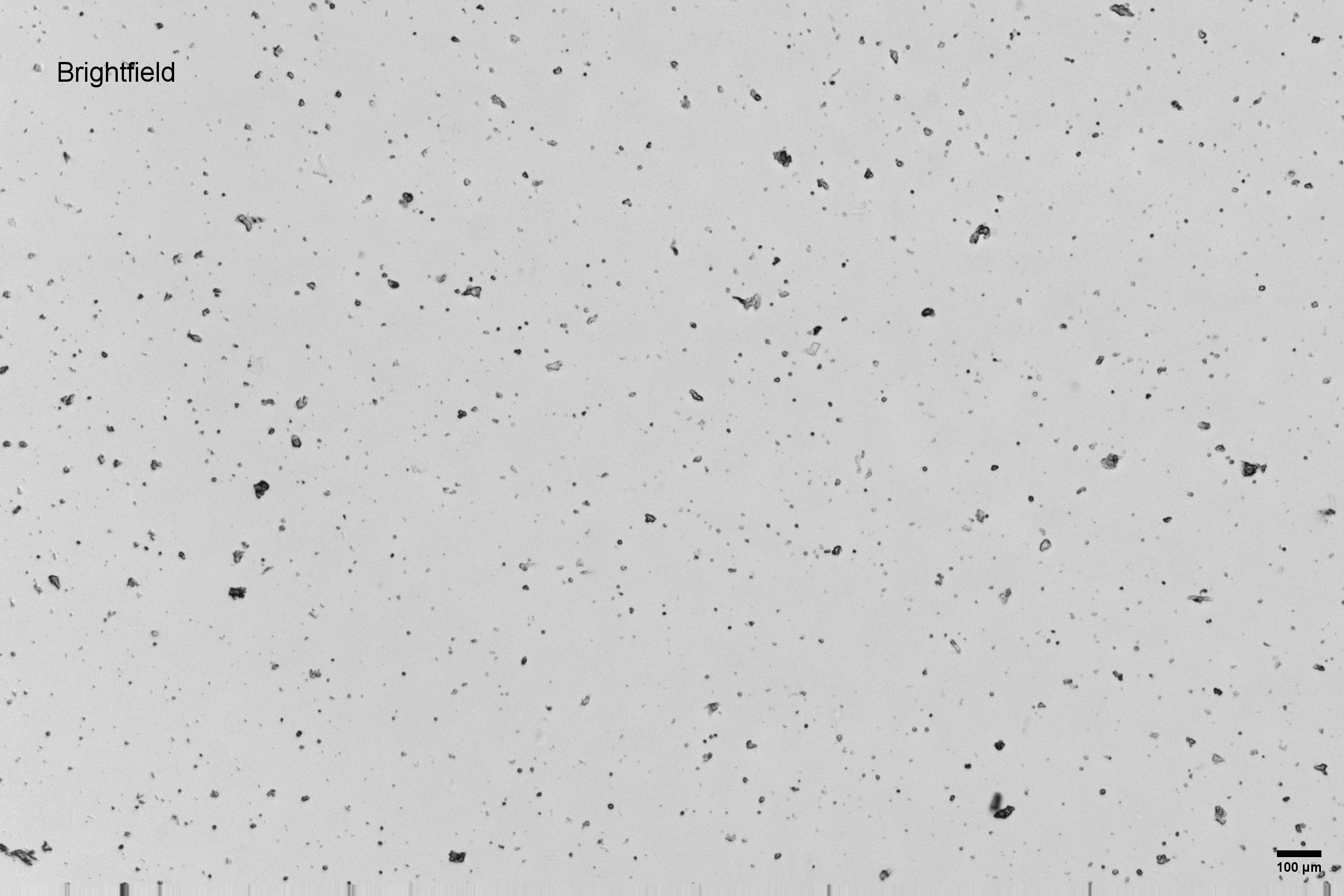 | 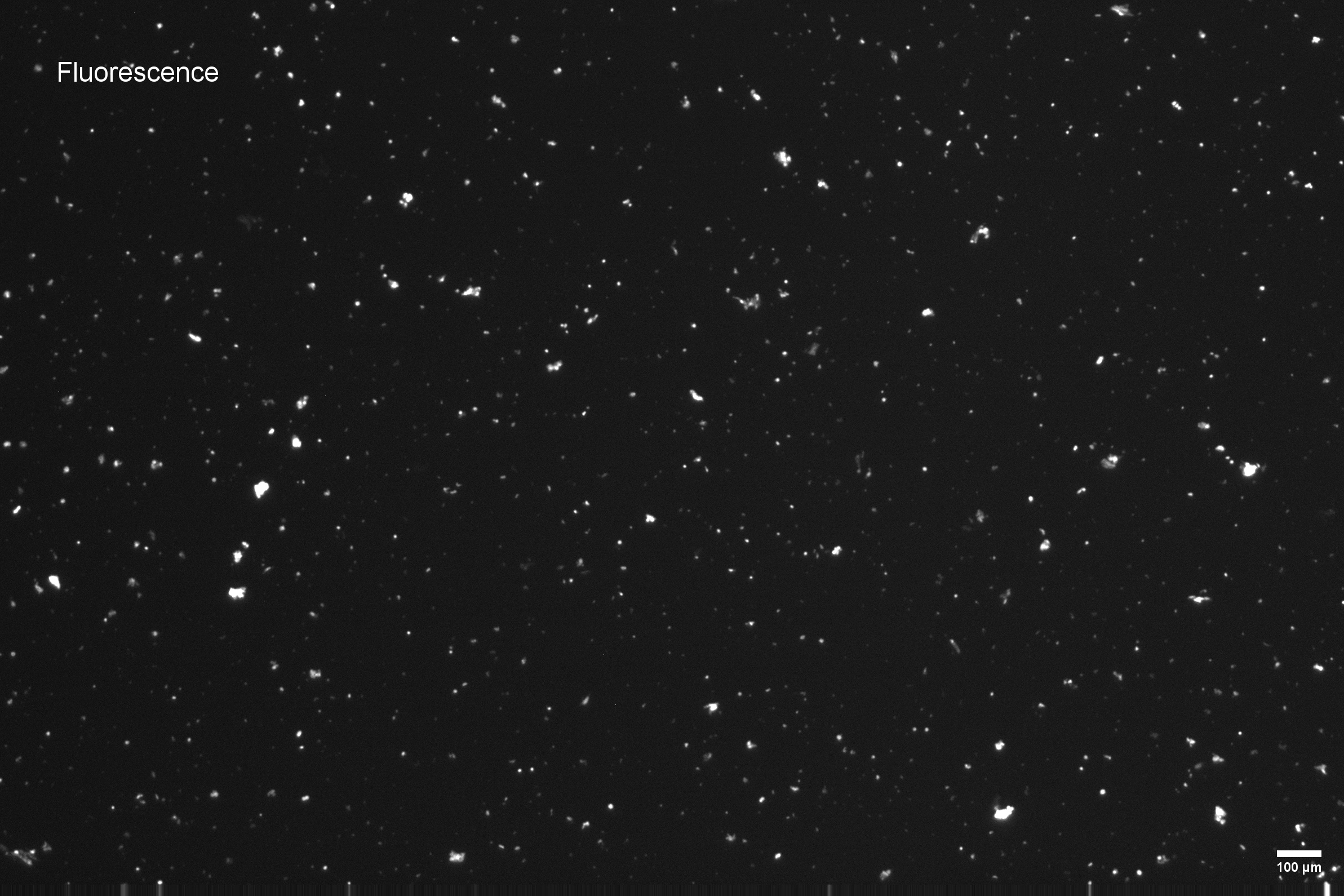 |
This dataset is licensed under the Creative Commons Attribution 4.0 International (CC BY 4.0) license. 10x citation guidelines available here.