Advancing CRISPR screens to build the "next human cell atlas": Cell type–specific gene function in neuronal cells

A new era of biological discovery is within reach thanks to the capabilities provided by recent advances in CRISPR screening technology. Having laid the groundwork to catalog the hosts of genes in the human genome, as well as the breadth of cell types in the human body, scientists are now able to define the functional roles of those genes and how gene function may change from cell to cell. Single cell CRISPR screening is at the leading edge of this movement, enabling comprehensive insights into cell type–specific gene function through sequencing and direct linkage of single guide RNAs to whole transcriptome gene expression effects in the same single cells. Read on to explore what’s now possible with this technology and how scientists are creating new atlases of gene function in the context of oxidative stress and neurodegenerative disease, for the first time, in human neurons.

The world has seen amazing scientific progress in the face of some of the most daunting tasks. The Human Genome Project began out of the lofty goal of sequencing the entire human genome in order to identify the genes that encode and enable life. In 2016, a global community of scientists initiated the Human Cell Atlas project, with the mission of characterizing every cell type in the human body to understand and advance human health. Today, scientists are beginning to envision a new kind of atlas—one that bridges both of its predecessors, gene and cell, to build a more comprehensive understanding of how genes function uniquely according to their cell type and, thus, what determines cellular heterogeneity beyond relative gene expression.
“Beyond gene expression, characterizing gene function in different cell types will be the next step toward building complete reference maps for the human body at the cellular level. [...] Parallel genetic screens across the full gamut of isogenic human cell types will uncover context-specific roles of human genes, leading to a deeper mechanistic understanding of how they control human biology and disease” (1).
There are 19,000–22,000 protein-coding genes in over 200 unique cell types in the human body, with more cells being discovered all the time (2, 3). If defining cell type–specific gene function across the breadth of these diverse genes and cells represents the next human cell atlas, robust genetic screening methods are urgently needed.
Advances in CRISPR screening technology are enabling scientists to begin to chip away at this huge task. In a recent study published in Nature Neuroscience, a team of researchers led by Dr. Ruilin Tianat of the University of California San Francisco (UCSF) leveraged pooled and single cell CRISPR screening to assess gene function in human induced pluripotent stem cell (iPSC)-derived neurons, representing the first genome-wide functional genomics assessment in this cell type (1). Their efforts have resulted in a new database called CRISPRbrain—an open source, cloud-based data sharing platform to catalog functional genomics screens in differentiated human cell types. They’ve started, of course, with brain cells, but the door is open for more researchers to share results and collaborate to continue building out the atlas.
In the sections below, explore the technology that is enabling these inroads into cell type–specific gene function and how, with this comprehensive information, scientists are able to uncover the genetic pathways controlling cellular responses in the context of pathology, even revealing potential therapeutic targets for drug development.
Behind the technology: Understanding pooled and single cell CRISPR screens
The UCSF team approached their experiment in two ways, beginning with pooled screens, which utilize a library of different CRISPR target guides in a single preparation, or pool, and which are delivered in bulk into cells of interest. As cells express those guides and CRISPR perturbations occur, the cell population will change. Cells expressing guides will become a smaller or larger part of the total population, indicating if the targeted genes are lethal, killing the cells, or growth advantageous, causing cells to proliferate.
Tianat et al. were specifically searching for the gene modulators of the neuronal response to chronic oxidative stress. Oxidative stress is often present in the context of neurodegenerative disease; therefore, clarifying how neurons respond to it represents an important step to better understand the underlying mechanisms of neurodegeneration and psychiatric disorders. They prepared genome-wide single-guide RNA libraries covering 19,000 protein-coding genes and used pooled CRISPR interference and activation (CRISPRi/a) screens in human iPSC-derived neurons to assess whether loss or gain of gene function, respectively, drove a toxic or protective effect.
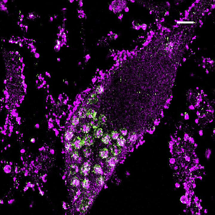
The screen revealed a number of perturbations with toxic knockdown phenotypes, including genes encoding superoxide dismutases SOD1 and SOD2, proteins that protect cells from oxidative stress (1). The research team noted that these proteins are not typically essential in other cell types, pointing to a particular need in neuronal populations and, therefore, a selective neuronal vulnerability to oxidative stress. They also observed toxic effects as a result of both knockdown and upregulation of several genes involved in protein homeostasis, suggesting expression levels for these genes are finely balanced for cell survival. Unexpectedly, knockdown of the lysosomal protein prosaposin (PSAP) also sensitized neurons to oxidative stress by driving production of lipofuscin, also known as “age pigment,” which accumulates in lysosomes over time and can cause iron buildup as well as cell death (4, 5). This last finding points to a novel role for PSAP in the neuronal response to oxidative stress and the larger narrative of neurodegeneration.
While very powerful, this pooled approach has some limitations as the final readout of gene function ultimately comes down to a simplified view of either cell death or cell survival. Gene expression content from living cells can be sequenced in a separate experiment to assess transcriptomic changes as a result of perturbation. But this and other stepwise aspects of pooled CRISPR screening can also make the effort to assess gene function in different cell populations logistically challenging, requiring labor-intensive parallel experiments that can take many weeks to complete.
Tian et al. noted similar challenges. Hundreds of the genes they had characterized in their pooled screen were associated with neurodegenerative diseases; however, they had a limited view of the function of those implicated genes. Commenting on this fact, the authors said:
“...Functional characterizations of these risk genes are largely lacking. Our CRISPRi and CRISPRa platforms provide a high-throughput approach to systematically interrogate gene function in human neurons. Beyond one-dimensional phenotypes, such as survival or fluorescent reporter levels, CRISPR perturbation can be coupled to single-cell RNA sequencing, using CROP-seq or Perturb-seq strategies to provide rich transcriptomic phenotypes” (1).
Indeed, innovations that have allowed the integration of high-throughput single cell sequencing and CRISPR screening technology represent a new approach with immense promise to advance the field of functional genomics.
The Chromium Single Cell CRISPR Screening assay from 10x Genomics is enabled by Feature Barcode technology, a system for barcoding biological analytes, such as cell surface proteins or single-guide RNAs (sgRNAs), with specialized oligonucleotide sequences that can trace them back to a cell of origin. See how this technology works in the context of single cell CRISPR screens by watching the video below:
With Feature Barcode technology, scientists can access sequencing information about the gene perturbation in an individual cell alongside whole transcriptome gene expression effects from that very same cell. Importantly, this can all be done within the same CRISPR screen, enriching the information collected from one experiment and streamlining parallel genetic screens of multiple gene perturbations in a population of cells. Together, this can allow scientists to obtain comprehensive gene function readouts beyond survival and a high-resolution view of perturbation effects across cell populations with greater efficiency and scale.
Narrowing in on neurodegenerative risk genes with single cell CRISPR screens
Looking to deepen their insights into the function of the neurodegenerative disease-associated genes they had identified in their pooled screen, the UCSF team then turned to single cell CRISPR screens using 10x Genomics Feature Barcode technology as a second approach to their experiment. They targeted 184 genes for CRISPRi and 100 genes for CRISPRa, representing a subset of disease-associated genes from their original library.
[Check out this infographic for a quick overview of the single cell CRISPR screening workflow.]
After sequencing their gene expression and CRISPR (Feature Barcode) libraries, linking perturbations directly to the transcriptomic effects in individual cells, the research team performed several different kinds of analysis. First, they identified differentially expressed genes (DEGs) resulting from each perturbation. This allowed them to identify functionally related genes whose perturbation yielded similar transcriptomic signatures. Next, weighted gene co-expression network analysis (WGCNA) allowed them to define genes that were co-regulated under different perturbations and build gene modules that could reveal more information about the relationships between those genes. In particular, the team identified a subset of genes with a previously uncharacterized role in neuronal fate regulation (1).
Three hypotheses for the role of NQO1
Single cell data also directed the research team to new hypotheses for underlying neurodegenerative disease mechanisms involving NQO1, a gene that encodes an oxidoreductase enzyme. NQO1 has been shown to have elevated expression and activity in the brains of patients with neurodegenerative disease and was previously thought to play a protective role against oxidative stress (1).
Instead of providing protection, however, overexpression of NQO1 in the pooled CRISPR screen had a negative effect on neuronal survival. Transcriptomic data from the single cell CRISPR screen provided insight into how NQO1 works to drive this toxicity phenotype. The team noted that overexpression of NQO1 was accompanied by increased expression of p53 and other p53 target genes. They also observed that NQO1 overexpression reduced expression of genes involved in axon development and cholesterol metabolism, which could contribute to cell death. Finally, they found that NQO1 overexpression also induced target genes of the transcription factor Nrf2. This was an unexpected result, as Nrf2 typically functions upstream of NQO1, regulating the expression of various antioxidant genes, including NQO1. This surprising feedback loop is all the more curious, as the Nrf2 pathway is also induced in some neurodegenerative diseases. Though there is more to be done to fully understand the relationship between Nrf2 and NQO1 overexpression, the team’s findings potentially reverse the current understanding of this gene pathway, suggesting it may contribute to disease rather than protect neuronal populations from oxidative stress (1).
Rich insights lay the foundation to build on CRISPRbrain
With the depth of information that researchers can access from single cell CRISPR screening approaches, there is great possibility for crucial insights into disease mechanisms and novel routes for therapeutic development—not only for neurodegenerative conditions but across all major classes of disease. And gene function itself is no longer bound to a simplified readout of cell death or survival. Rather, technological innovation in CRISPR screening is allowing our experiments to match the true complexity of biology, resolving the transcriptomic effects of perturbation at single cell resolution for a rich picture of gene function in specific cell populations. The tools in place—and the experiment modeled through this team's efforts to generate CRISPRbrain, an atlas of gene function across cell types—scientists have what they need to add new experiments, new cell types, and new knowledge to our collective understanding of functional genomics.
References:
- Tian R, et al. Genome-wide CRISPRi/a screens in human neurons link lysosomal failure to ferroptosis. Nat Neurosci 24: 1020–1034 (2021). doi: 10.1038/s41593-021-00862-0
- Willyard C. New human gene tally reignites debate. Nature 558: 354–355 (2018). doi: 10.1038/d41586-018-05462-w
- Yong E. A Google Maps for the Human Body. The Atlantic. October 14, 2016.
- Terman A and Brunk UT. Lipofuscin: mechanisms of formation and increase with age. APMIS 106: 265–76 (1998). doi: 10.1111/j.1699-0463.1998.tb01346.x.
- Li J, et al. Ferroptosis: past, present and future. Cell Death Dis 11: 88 (2020). doi: 10.1038/s41419-020-2298-2.