Dr. Mats Nilsson shares his thoughts on the past, present, and future of spatial biology
Impact at a glance: In this article Dr. Mats Nilsson, a pioneer and leading expert of spatial methods, highlights how integral spatial biology will be in the near future, particularly for neuroscience and cancer biology. He highlights the power of the Xenium platform in these endeavors given it “produces data that’s just on a little bit of a different level” and his belief that single cell spatial data will become a required component of single cell work.
“We’re convinced that spatial biology is the future. We've been convinced about that for a couple of decades. I want to be working on methods to try to address molecular profiling in tissues.” –Mats Nilsson, PhD, Professor, Stockholm University, Sweden
New advances in science are often heralded by novel technologies. It makes sense, after all: we’re limited not by our imaginations, but available tools. And one such researcher, Dr. Mats Nilsson at Stockholm University, has spent his career dreaming up the tools to make those limitations an afterthought.
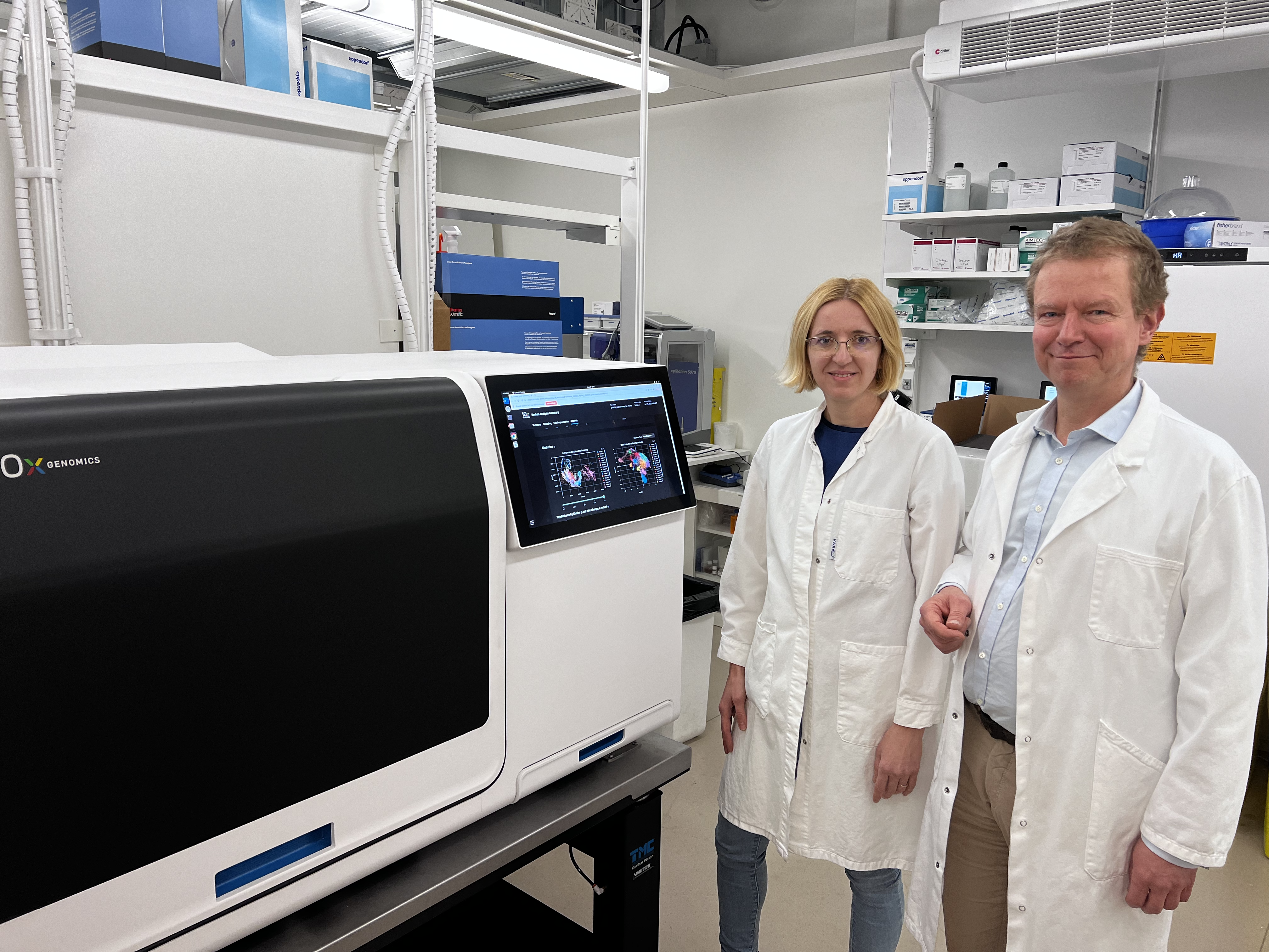
His work was instrumental in helping bring our Xenium In Situ platform to life. So of course we jumped at the opportunity to interview both him and the head of the in situ sequencing unit at Science for Life Laboratory—Katarina Tiklova, PhD—about where spatial biology has been and where they think it’s going.

Thank you so much for agreeing to talk with us. So, to kick us off: what got you started with spatial biology, and where do you see it going?
Dr. Nilsson: We're convinced that spatial biology is the future. We've been convinced of that for a couple of decades. I want to be working on methods to try to address molecular profiling in tissues. And, as a result, we developed in situ sequencing technology and chemistry, which is one of the foundations the Xenium instrument is built upon.
We are great believers in the importance of addressing biology on an organ level, and it's really the logical step to take after the single cell revolution. I would say that the single cell revolution made the spatial revolution useful because the technologies were out there, but we couldn't really use them in an efficient way.
Before, we understood the molecular compositions of cell populations. Then it became obvious we needed their spatial architecture in the tissue. That’s the real objective, I think, of modern cell biology, and this is sort of where we come from.
How are you using Xenium and spatial biology methods in your lab?
Dr. Nilsson: We apply the technologies with our in-house developed chemistries and also with Xenium. So we run both in parallel. We're very happy with the Xenium platform because it generates the same kind of data we managed to generate before, but so much better and so much easier. It's really a dream come true that there’s a machine that generates this fantastic data.
We've been doing basic fundamental cell atlasing projects to try to understand the spatial locations of different cell populations identified by single cell RNA sequencing.
And then I have a particular interest in trying to characterize and understand the most complex tissues of them all: tumors and cancer tissues, because they're really not reproducible. So every piece of tissue is unique, and, of course, that’s a challenge, but I think that's also where we can expect that this spatial approach will have the greatest clinical impact going forward.
Dr. Tiklova: For me, I was working with single cell sequencing using a model for Parkinson’s disease. We characterized a lot of subpopulations, then we needed to go back and identify them in space, so that was the start of spatial biology for me. It was around that time that I got in touch with Mats.
We had all this data, but we needed to look at the cells in space, so we used what was actually the first version of in situ sequencing. It was still sequencing by ligation, which I was using for my postdoc project. We managed to publish it, in collaboration with Mats at the time. Then, when I finished my postdoc, there was a position in the Science for Life Laboratory spatial biology core facility and I applied for that.
Having experience with manual in situ methods, we really appreciate the Xenium instrument. It saves a lot of time, and it produces data that’s just on a little bit of a different level.
So, a two-part question given a couple points you made. First, you commented that Xenium, “produces data that's just on a little bit of a different level." Can you expand on this a little? Do you mean sensitivity, specificity, reliability, or something else?'
Second, you said that you were very happy with Xenium because it, “generates the same kind of data you generated before, but better and easier.” What makes it better and easier? Is it the workflow, the bioinformatics tools, and/or something else?
Dr. Tiklova: For the first part, we meant that the Xenium workflow is more streamlined, more automated, and takes issues such as highly expressed genes, cell segmentation, etc. into consideration. This produces more specific and reliable data, with more RNA transcripts resolved per cell, compared to our homemade chemistry.
For your second question, automated imaging and decoding saves a considerable amount of time and leads to more consistent and reliable data. This is crucial in handling large datasets. The integrated bioinformatics tools play an important role in making the complex data accessible to biologists without extensive bioinformatics knowledge. Also, tailoring the number of probes to the expression level of a gene provides better results.
Touching on panels: what are your thoughts on panel design? There’s a lot of debate right now about how many genes should be in a panel, with some favoring thousands of genes. Where do you see very high-plex panels being more beneficial to customers, and where do you see more focused panels being most useful?
Dr. Nilsson: Explorative studies will benefit from more genes, there’s no doubt about it. Genomics is very powerful because you see everything at the transcriptomic level; you don't need a particular idea or hypothesis before you test your samples, you just look at everything. The closer you come to specific questions, maybe diagnostic questions, the fewer genes you’d like to target.
Dr. Tiklova: I believe the same thing. If you make it too complex, it will be very difficult to interpret things. It has to go fast, and it has to have fast answers. That’s why I believe that Xenium’s targeted approach will succeed in the clinic because you don’t need all the genes.
We think that a lot of people are now realizing that spatial biology is the future and where they’ll need to go. Has there been any skepticism? How do you think that’s progressed as the technologies have gotten easier and more accessible? And how do you validate your single cell spatial imaging experiments?
Dr. Nilsson: People connect with us because they want to do spatial, and I don't think there's any real skepticism about spatial. What would you say, Katarina?
Dr. Tiklova: I think everyone understands that’s where you need to go now. You can’t only know what the cells are because the location is even more important than the whole expression. So now I don't think we really have to convince people, but it's still a part of a continuum: a lot of people would be interested, but the cost of spatial experiments is still pretty high at this point. If you go back to the single cell sequencing era, it was the same. Everything cost so much, you had to limit how many cells you could sequence.
Dr. Nilsson: So the cost per data point will be the major driver going forward, and it will happen: with your company making this so much more efficient and scaling up, things will become cheaper. It will open new kinds of studies and new users, and the volumes of samples will increase with the drop in price.
Dr. Tiklova: And for that last point, we typically validate our single cell spatial imaging data using other available datasets like the Allen Brain Atlas, antibody staining, and similar methods.
Early last decade, we started seeing single cell sequencing lead to an explosion of papers that characterized individual cell types and identified disease-associated cell types. Then, spatial transcriptomics came in, and we had an explosion of papers focused on anatomical characterization and spatial context of these changes. Given what Xenium is capable of, what sort of themes do you think we'll see going forward?
Dr. Tiklova: At the beginning, I think there will be a focus around tissues that are easy to analyze since you have to address technical challenges for some tissue types (such as bone). I think there will be a lot of papers focused on brain tissue as well, given that isolating neurons for single cell sequencing is more challenging. I think there will be a boom in the cancer field because you have so much variation in each individual tumor.
Dr. Nilsson: I expect to see a lot of cell atlasing and characterizing of human disease samples that are geared towards understanding their cellular compositions. And, I think that single cell spatial imaging will be a required experiment in the future. I don’t think you’ll be able to publish single cell data without the Xenium experiment to back.
Thank you both very much for your time today!
Dr. Nilsson: You’re welcome; have a nice day.
This interview has been edited for length and clarity.
See how Dr. Nilsson is driving spatial biology forward with technological innovations on his lab’s website, and explore how our Xenium In Situ platform is ushering in a new era of spatial biology (as well as our roadmap of upcoming features, including high-plex panels and enhanced cell segmentation).
