To be a meningeal fibroblast: scRNA-seq transforms our view of cellular heterogeneity in the meninges

Dr. Julie Siegenthaler, Associate Professor in the Department of Pediatrics, Section of Development Biology, at the University of Colorado, has studied the meninges for years. Now, she is filling a knowledge gap in the neuroscience community’s understanding of embryonic meningeal fibroblast heterogeneity, development, and function, with the help of single cell RNA-sequencing. These findings have the potential to illuminate the role of central nervous system (CNS) vasculature on the development of the embryonic brain and crucial protective structures like the blood-cerebrospinal fluid barrier.
When it comes to understanding central nervous system (CNS) development, health, and disease, researchers need to think outside of the box. Sometimes that leads them to structures outside of the brain. The meninges are composed of three membrane layers, including the dura, arachnoid, and pial layers, which encase the CNS from the earliest stages of embryonic development and serve as a protective covering later in adulthood. These layers contain various cell types, including meningeal fibroblasts and immune cells, and are integrated with a network of blood and lymphatic vessels that deliver oxygen, nutrients, and developmental signals to the CNS.
![The meninges of the central nervous system. CREDIT: SVG by Mysid, original by SEER Development Team [1], Jmarchn - Vectorized in Inkscape by Mysid, based on work by SEER Development Team. (CC BY-SA 3.0).](https://cdn.10xgenomics.com/image/upload/c_limit,f_auto,h_510,q_auto,w_680/v1600272455/blog/2560px-Meninges-en.svg.png)
Dysregulation in meningeal fibroblasts is also known to underlie CNS developmental defects. A previous study of Foxc1 mutant mice revealed that loss of Rdh10- and Raldh2-expressing cells in the cerebral meninges, and subsequent loss of meninges-derived retinoic acid signaling, correlated with severe neocortical defects (1). Similar Foxc1 mutations have been observed in genetic studies of human cerebellar malformation, including Dandy-Walker malformation (2), again pointing to the key role of the meninges in successful brain development.
For these reasons, Dr. Julie Siegenthaler, Associate Professor at the University of Colorado, and her research colleagues in the Siegenthaler Lab, are determined to understand the contribution of non-neuronal structures, like the meninges, to CNS development and function.
“We’re interested in the vasculature because it's essential for brain metabolism, but it also represents an area where the central nervous system can access signals that come from the periphery … and that regulates brain function. How does the CNS vasculature acquire properties that enable the CNS to communicate with the periphery, but then also protect it? This is set up developmentally in the formation of the tight junctions between the endothelial cells, the acquisition of pericytes and astrocytic end feet, and the establishment of the neurovascular unit. All this is done, at least in mice, within a couple of weeks of age. So this is all absolutely essential to brain development and function, and these structures are heavily dysregulated in a variety of diseases.”
Components of the meninges, including meningeal fibroblasts, are also crucial sources of peripheral signaling during the early stages of neural development. As Dr. Siegenthaler notes, “the vasculature and meninges represent these really essential support structures for brain development and function,” however they are not as well characterized as the breadth of neurons and cell types represented in the brain and spinal cord. “The fibroblasts of the meninges had never been profiled … they typically just get labeled with very bland fibroblast markers when we really don’t understand what it means to be a pia or an arachnoid or a dura cell.”
A new perspective on meningeal fibroblast diversity
In an effort to explore the true diversity of meningeal fibroblasts, and uncover their specialized roles in the developing CNS, Dr. Siegenthaler and her colleagues turned to single cell RNA-sequencing. “This was really a big gap in knowledge, and this is what single cell technology is perfect for. We’ve done bulk RNA-seq on the meningeal fibroblasts, sorting them based on expression of Collagen1a1-GFP. Essentially, you just get a blend of … mixed cell populations, and it's very difficult to make any type of interpretation.”
With single cell resolution, however, Dr. Siegenthaler’s group was able to develop distinct molecular profiles of different meningeal fibroblast populations derived from E14 mouse forebrains. Using scRNA-seq, they captured and sequenced the transcriptomes of 8,581 GFP+ FACs-sorted meningeal fibroblast cells and grouped those populations into six principal clusters based on gene expression. Further analysis segregated the meningeal subclusters into three main branches that correspond to the pial, arachnoid, and dura classifications.
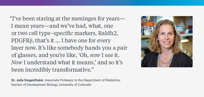
These insights into the distinct cellular markers between meningeal layers represent a breakthrough in meningeal fibroblast research. As Dr. Siegenthaler expressed, “I’ve been staring at the meninges for years—I mean years—and we've had, what, one or two cell type–specific markers, Raldh2, PDGFRβ, that's it. That's all we've had for years. ZIC1 isn’t even very good because it would hit all of them. I have one for every layer now. It's like somebody hands you a pair of glasses, and you’re like, ‘Oh, now I see it. Now I understand what it means,’ and so it's been incredibly transformative.”
Importantly, this research revealed new cellular markers for arachnoid barrier cells, a population within the meninges that forms tight junctions. “This becomes a tight-junction containing layer that exists within the meninges and is part of the barrier system that prevents cells and molecules from getting into the cerebrospinal fluid.” Once a “wildly under-studied structure,” the blood-CSF barrier is becoming less of a mystery, as Dr. Siegenthaler’s team continues to explore its cellular composition and differentiate cellular populations at the transcriptional level. “We had no markers for these arachnoid barrier cells… Now we know what they express. We can label them. We can study them. We know the signaling pathways. Based on the single cell data, we made predictions about signaling pathways that are important for their development. And all of this came out because we were able to look at and pull these cells out and see, ‘Oh, these guys represent a new population of cells. What did they express? How are they different?’”
Insights into the layer-specific and cell type–specific markers opened up even more possibilities of discovery for Dr. Siegenthaler’s team. Further analysis via in situ gene expression in E14 brain tissue sections revealed regionalized expression patterns, meaning that subclusters with distinct transcriptional profiles identified within the meningeal layers correlated with meninges adjacent to specific brain regions. Single cell meningeal fibroblast data also pointed to unique developmental functions among subpopulations of the embryonic meninges. For example, Dr. Siegenthaler’s group noted that pial subclusters had enriched expression of extracellular matrix components, suggesting pial fibroblasts play the biggest role in helping to form the pial basement membrane, which divides the brain parenchyma and the meninges. They also identified a unique fibroblast subpopulation in the pial layer with heightened expression of Crym and Serpine2, the latter of which is a known factor in inhibiting angiogenesis. Consequently, they concluded that this subpopulation may have region-specific functions related to the regulation of meninges-specific structures like blood vasculature (3).
Opening new doors for other neuroscience researchers
With the knowledge they’ve gained from their own single cell study of the meninges, Dr. Siegenthaler and her team have been able to support other researchers interested in detailed annotations of meningeal fibroblasts. “We have gone back and analyzed other people’s data… This is actually really helpful. Instead of, basically, fibroblasts, just an anomalous cluster in these big single cell brain annotations, people can actually annotate those clusters and say, ‘Oh, yeah, these are the arachnoid barrier cells. These are the pial cells. These are the non-arachnoid barrier cells. These are the dural fibroblasts. Or perivascular fibroblasts.’ That enriches their dataset.”
Explore these cellular insights in the Siegenthaler Lab E14 Meningeal Fibroblast Single Cell RNA-seq Data Set.
In addition to sharing data and sharpening insights from past studies, Dr. Siegenthaler has a number of tips for researchers who are getting started with single cell analysis. “You’ve got to have the right question,” and if you don’t yet, or if you’re not sure where to start, find out if “somebody has done it already and the data is publicly available. The first thing to do is go and analyze other people's data.” Dr. Siegenthaler noted that analyzing other people’s data is also a powerful training mechanism if you’re looking for more bioinformatics practice. Finally, she recommends pouring into some helpful single cell reviews: “I think there are a lot of great reviews on single cell technology that I would highly encourage people to read, particularly about sample prep—I think that’s essential. Are you sorting cells? What kind of digestion methods are you using, and how is that going to affect the transcriptome?”
Thank you Dr. Siegenthaler for sharing your tips and insights! We look forward to hearing from more scientists for whom single cell analysis is unlocking the hidden cellular heterogeneity that has kept researchers guessing for years.
References
- J Siegenthaler et al., Retinoic Acid from the Meninges Regulates Cortical Neuron Generation. Cell. 139, 597–609 (2009).
- KA Aldinger et al., FOXC1 is required for normal cerebellar development and is a major contributor to chromosome 6p25.3 Dandy-Walker malformation. Nat. Genet. 41, 1037–1042 (2009).
- J DeSisto et al., Single-Cell Transcriptomic Analyses of the Developing Meninges Reveal Meningeal Fibroblast Diversity and Function. Develop. Cell. 54, 43–59.e4 (2020).
This article contains a discussion of research conducted in the laboratory of Dr. Julie Siegenthaler. View and opinions do not constitute endorsement or promotion of 10x Genomics, Inc. or any of its products.