Translational applications of single cell and spatial sequencing at the EMEA Multiomics Discovery Symposium

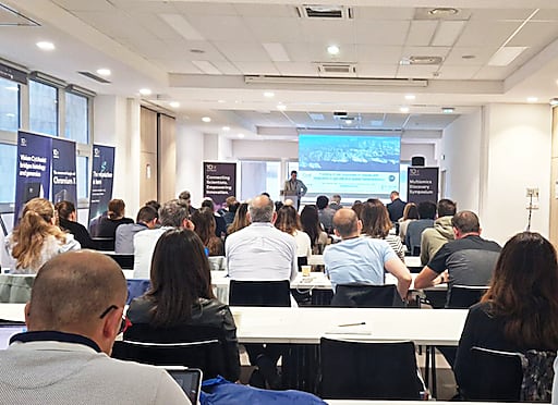
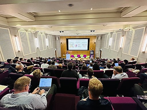
Researchers around the world are resolving the underlying mechanisms of disease, from cancer to allergies, in order to develop better therapeutic approaches and advance human health. This shared effort brought together a global community of leading scientists and 10x Genomics experts at the recent Multiomics Discovery Symposium—held consecutively at three European cities, Marseille, London, and Heidelberg—to explore applications of single cell and spatial technology to translational research. Their discoveries reflect the potential of high-resolution, multidimensional analytical approaches to match the complexity of disease and define the cellular and molecular features that may serve as biomarkers for predictive diagnosis, roadblocks to more effective therapies, or the key to understanding successful therapeutic mechanisms of action.
Presentations highlighted at these global gatherings also reflect the importance of sharing knowledge as a scientific community, across disease areas and applications of single cell and spatial technology. Whether it’s best practices for challenging sample prep or tips for data analysis, one scientist’s experiences can galvanize another’s next experiment.
Keep reading to find translational research highlights from each of the three cities (including highlights on COPD lung cell atlases, allergen immunotherapy, and central nervous system tumors), as well as on-demand presentations for sample prep and data analysis best practices across the 10x Genomics suite of technologies—Chromium, Visium, and Xenium. Watch now →
Healthy and pathological human lung cell atlases build foundation to understand COPD
The lungs are complex organs. They contain around 40 billion cells, which are grouped into an estimated 60 distinct cell types. Specifically, the Human Lung Cell Atlas currently reports having profiled and defined the anatomical locations of 58 cell populations in the human lung, including 41 of 45 previously known cell types or subtypes and 14 new additions (1). Yet the cellular microenvironments of the lung can become even more complex in the context of inflammation and disease.
Chronic obstructive pulmonary disease (COPD), a lung disease that restricts airflow and impairs breathing, is the third leading cause of death worldwide (2). Dr. Pascal Barbry, research director at the French National Center for Scientific Research, and a speaker at our Marseille Multiomic Discovery Symposium, uses single cell RNA-sequencing to deeply characterize the cellular networks involved in COPD and build cellular atlases of both healthy and pathological human lung tissue to understand how cellular context affects gene expression.
Barbry presented findings from his group’s recent study, which leveraged single nuclei RNA-seq to profile over 380,000 cells from 109 samples, taken from 10 early-stage COPD patients and 12 healthy controls between 50–60 years old. This revealed 48 distinct cell identities, with two cell types, goblet cells and multiciliated cells, showing a subset of genes known to be upregulated by smoking that were not found in healthy controls. These findings may represent a first step towards defining cellular mechanisms of disease. They also observed different gene expression signatures in samples taken from various parts of the airway, such as between the nasal tissue or the tracheobronchial tissue, suggesting pathology manifested distinctly among unique lung regions.
These kinds of analyses can yield even more specific, confident results through scale. Barbry and his team compiled single cell data from other labs to generate a human lung cell atlas core, which included 2.2 million cells from 444 individuals. Analysis of this massive dataset allowed them to define over 60 cell types and states from lung airways and parenchyma, as well as improve cellular annotations of rare cell types that could have been incorrectly labeled in smaller datasets.
Like the theme of the larger Multiomics Discovery Symposium series, this core dataset reflects the scientific advancements that are possible through shared knowledge, collaboration, and powerful insights from high-resolution, high-throughput tools.
Exploring the potential of allergen immunotherapy for seasonal hay fever
Dr. Janice Layhadi, research associate at Imperial College London, told us in the London series of the Multiomics Discovery Symposium that over 500 million people worldwide suffer from allergic rhinitis (AR), colloquially known as seasonal hay fever. Unfortunately, common antihistamine or corticosteroid treatments for AR can only provide temporary relief—or no relief at all for 20% of patients.
But there’s another option for some patients: allergen immunotherapy (AIT), a disease-modifying treatment with potential long-term clinical benefits administered through subcutaneous injection or sublingually (under the tongue). Layhadi spearheaded research using single cell immune profiling and cell surface protein analysis to characterize the immune cell populations that may be responsible for the beneficial effects of AIT, focusing on a novel regulatory subset of innate lymphoid cells (ILCs). One specific class of ILCs—ILC2—showed the potential to produce interleukin 10 (IL-10) after exposure to grass pollen in vitro. IL-10 acted as an immunomodulatory cytokine, dampening the levels of another cytokine (IL-13) typically elevated in individuals with grass pollen allergies. Single cell phenotyping further distinguished an IL-10+KLRG1+ ILC population from T regulatory cells, confirming it was a novel, distinct cell type that played a role in controlling immune responses to allergens.
In a time-course clinical trial of allergen immunotherapy, Layhadi also used single cell immune profiling to demonstrate that patients treated with AIT had a much higher percentage of IL-10+ ILC2 clones compared to those treated with the placebo, confirming that ILCs from the treated cohort had a higher capacity to become IL-10 producers as a result of immunotherapy.
True to the goals of translational research, this single cell multiomic analysis of clinical patient samples is providing strong evidence to support a mechanistic understanding of the benefits of allergen immunotherapy. Layhadi’s ongoing clinical trial collaboration with a pharmaceutical company that has developed a chemically modified allergen (called an allergoid) that can be used in grass pollen immunotherapy also holds promise for continuous improvement of allergen immunotherapies for patients.
Building single cell and spatial maps of meningioma and glioblastoma
The tumor microenvironment holds both cellular and spatially resolved clues to cancer progression; therefore, merging these two worlds may produce the clearest map of tumor heterogeneity and the cellular relationships within and across tumors, infiltration zones, and healthy bordering tissues.
During our Heidelberg series of the Multiomics Discovery Symposium, Dr. Natalie Berghaus, postdoctoral researcher at the German Cancer Research Center (DKFZ) of Heidelberg University Hospital, discussed her team’s research into two central nervous system (CNS) cancers, meningioma and glioblastoma.
They used single cell RNA-seq to evaluate the heterogeneity of a representative cohort of human meningioma tumors, noting prominent cell types, including a cluster of macrophages reflected across all samples. Diving deeper into the differences between macrophage populations from different tumor grades—which were determined by the established WHO classification system and DNA methylation classification system—showed a decreasing proportion of infiltrating macrophages as tumor grades got higher. Patient correlative data confirmed that individuals with tumors containing lower proportions of infiltrating cells had shorter periods of progression-free survival, indicating that macrophage infiltration could serve as a cellular biomarker for prognostic or diagnostic purposes.
Though meningioma is a more common CNS tumor, glioblastoma is a more aggressive, heterogeneous cancer that has a diffuse growth pattern and no immunohistochemistry markers, making it difficult to not only diagnose, but also define the borders of the tumor and infiltration zones. This led Bergaus’ team to map known chromosome 7 gain events and chromosome 10 loss events in FFPE tumor tissue sections using Visium Spatial Gene Expression as a means to discriminate tumor regions. They also used an existing single nuclei dataset to deconvolve spatial data from glioblastoma samples, which allowed them to annotate specific cell types in a spatially resolved manner. Together, this revealed clear tumor regions, infiltration zones, and necrosis areas within tissue sections, as well as localization patterns of neurons and oligodendrocytes. Bergaus confirmed that this high-resolution spatial CNV analysis was possible even in small punches of tissue, marking a possibly transformative jump in molecular analysis capabilities for both challenging and precious clinical samples.
This was only a small sample of the great research shared at the Multiomics Discovery Symposium series. You can find more on-demand presentations highlighting translational research or experimental tips and tricks here →
Want to learn more about the technology discussed in this blog? Find resources for Chromium Single Cell Gene Expression, Single Cell Immune Profiling, and Visium Spatial Gene Expression.
Finally, a big shout out to our amazing customers and 10x Genomics experts who make these global meetings possible! Find a User Group Meeting (UGM) in your area →

References: