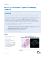
The Visium CytAssist is designed to analyze tissue sections placed on blank slides. Before running a Visium CytAssist assay, a high resolution image is captured with a microscope that is combined with the image taken by the instrument to overlay gene expression data. Successful NGS data and protein visualization are highly dependent on good imaging practices. This Technical Note provides hardware recommendations, general image acquisition and analysis guidelines, and examples of images that are suitable for downstream analysis with Space Ranger. Individual results may vary depending on the specific imaging system, and/or sample characteristics.
This Technical Note applies to the following CytAssist assays and sample types:
-
Visium CytAssist Spatial Gene Expression
- Hematoxylin & Eosin (H&E) stained formalin fixed & paraffin embedded (FFPE), fresh frozen & OCT embedded (FF), and fixed frozen & OCT embedded (FxF) tissue sections Immunofluorescence (IF) stained FFPE tissue sections
-
Visium CytAssist Spatial Gene and Protein Expression
- H&E and IF stained FFPE tissue sections