Breaking boundaries in single cell and spatial discovery: 10x Genomics symposium highlights
Berlin, March 5, 2024 - The 10x Genomics Single Cell & Spatial Discovery Symposium in Berlin exceeded all expectations, transforming from its original plan as a user meeting with 60 expected attendees into a vibrant symposium that drew over 200 participants. The symposium provided a stage for users of 10x Genomics technology to share their experiences as well as a forum for the company to unveil new products, address expectations, and explore emerging requests from the scientific community.
In the sections below, you’ll find an overview of the key insights from this event, including featured presentations from customer speakers:
- Benchmarking single cell 3’, Flex, and Xenium
- Flex and Xenium reveal gene signatures in Crohn's disease
- Xenium offers insights into the dynamics of Herpes infection
- Comparing single cell 3’ and Flex
- Clinical research capabilities from a 10x Genomics Certified Service Provider
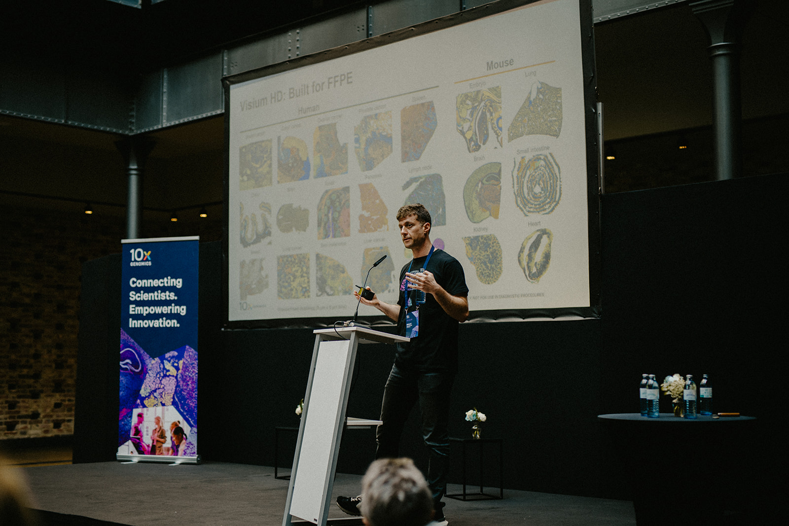
- FFPE-compatible Flex and Visium assays define the evolution of non-small cell lung cancer
I. The users
1. Thomas Conrad, PhD
Benchmarking Single Cell Gene Expression kits and Xenium Analyzer
Dr. Thomas Conrad, head of the Single Cell Technologies Team at the Max Delbrück Center and BIH/Charité, showcased the widespread adoption of 10x Genomics technology within his facility. Impressively, more than 80% of their research endeavors rely on the robust and versatile capabilities of 10x Genomics technology. During the meeting, he presented the results of two benchmarking experiments, both using organoid models.
The first experiment compared the Chromium Single Cell 3' Gene Expression kit (3’ Gene Expression kit) and the Chromium Single Cell Gene Expression Flex kit (Flex kit), with the conclusion that Flex exhibited higher sensitivity and specificity.
The second benchmarking study highlighted Conrad's team's experience with the CosMx SMI from NanoString and the Xenium Analyzer from 10x Genomics. He demonstrated the high-throughput power of Xenium, noting the significant advantage of reduced processing time—from 2 weeks to 2 days—for tissue microarray organoids. He also discussed his experience with the newly launched Xenium Cell Segmentation kit, which offers improved cell-type delineation and transcript assignment, a particularly beneficial feature for challenging samples like bone marrow and heart tissue. The team's dedication to advancing spatial omics analysis is evident in their publication on the VoltRon method.
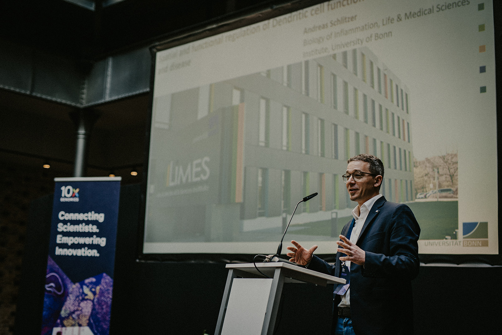
2. Andreas Schlitzer, PhD
Unraveling inflammation in Crohn's disease with Single Cell Gene Expression Flex and Xenium Analyzer
Dr. Andreas Schlitzer, Chair of the Biology of Inflammation Life and Medical Sciences Institute at the University of Bonn, explored the intricate role of mature dendritic cells enriched in immunoregulatory molecules (mregDC) in inflammation, particularly in Crohn's disease. Utilizing the Flex kit, Dr. Schlitzer's team analyzed gene expression across various tissues, including lymph nodes, adipose tissue, synovial fluid, and peripheral blood monocytes, comparing healthy individuals with those experiencing short-term or long-term infections. Their study unveiled a distinct gene signature present in chronic inflammation, particularly evident in lymph nodes.
The team did more research using the Xenium assay in the lymph nodes of people with refractory Crohn's disease and found that samples from these individuals had more mregDC cells and a granuloma (a non-cancerous cluster of white blood cells and other tissue often seen in inflammatory diseases like Crohn’s) that was mostly made up of macrophages. The identification of an elevated number of inflammatory mregDC cells emerged as a predictive marker for susceptibility to chronic inflammation. This groundbreaking finding also offers potential stratification for more effective Crohn's disease treatments.
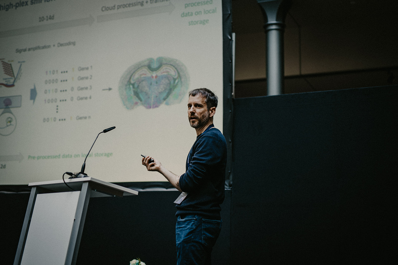
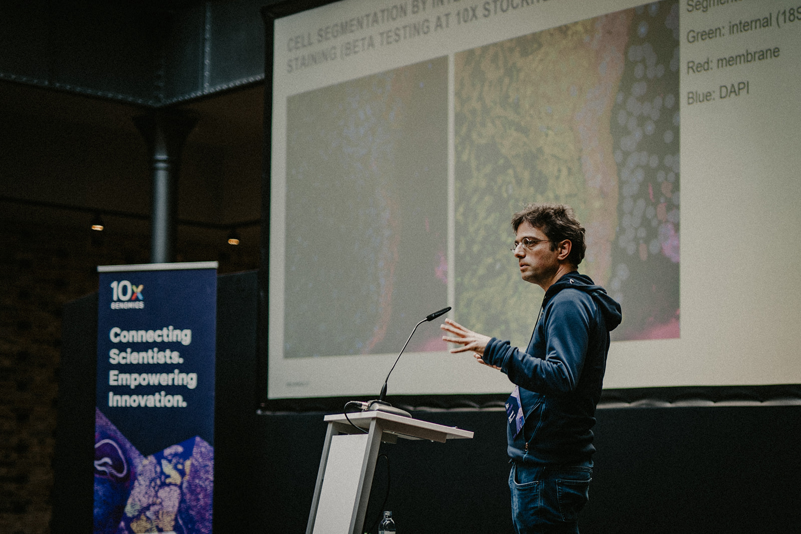
3. Emanuel Wyler, PhD
Understanding herpes simplex virus infection with Xenium technology
Dr. Emanuel Wyler, a scientist at the Max Delbrück Center, unveiled groundbreaking research centered on skin organoid models for exploring herpes simplex virus (HSV) infection. This virus impacts 80% of the global population, posing particular risks for newborns and immunocompromised individuals. Despite current treatments, the quest for a definitive cure or vaccination remains an ongoing challenge.
Utilizing Xenium technology, Dr. Wyler's team integrated a custom gene panel to localize HSV within the organoid sections. Additionally, they designed probes for other viruses, such as papilloma, influenza, and the flaming virus, for parallel experiments. Although they encountered hurdles, particularly with sequencing variable regions for the flaming virus, the overall spatial outcomes were excellent. Early access to the Xenium Cell Segmentation kit significantly improved result definition compared to sections where it was absent.
Further analysis of the HSV infection samples with the 3’ Gene Expression kit revealed that the virus prompts keratinocytes to express TNF-alpha. This, in turn, induces surrounding cells to express the inflammatory factor and alter their transcriptome, facilitating the virus's spread within the organoids. The 3' Gene Expression kit played a crucial role in analyzing the transcriptome of individual skin organoid cells, despite the challenges encountered in keratinocyte dissociation. This research provides crucial insights into the dynamics of HSV infection, offering potential avenues for therapeutic advancements.
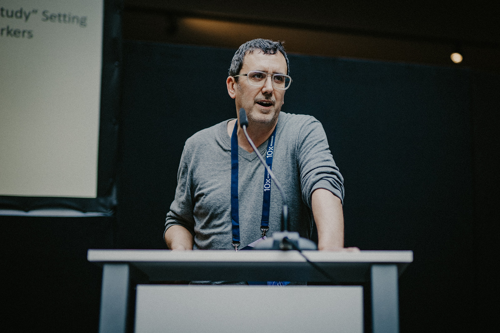
4. Markus Morkel, PhD
3’ Gene Expression kit or Flex kit?
Dr. Markus Morkel, leader of the Laboratory of Molecular Tumor Pathology at the Bioportal Single Cells, Institute of Pathology, Charité, University of Berlin, is a key contributor to the Multi Omic Spatial Atlas In Cancer (MOSAIC) consortium. As part of this project, his team will be responsible for processing up to 7,000 paraffin-embedded (FFPE) samples, only feasible through the Flex kit. While acknowledging the kit's remarkable value and flexibility, Dr. Morkel also emphasized to users that their technology choices should align with their specific research objectives.
His comparative analysis revealed striking similarities in data obtained from parallel experiments using the 3’ Gene Expression and Flex kits. Notably, the FFPE samples demonstrated a more robust representation of cell diversity. Addressing gene expression signatures, Dr. Morkel found similarities, yet certain genes exhibited differential representation in each kit. While praising the Flex kit for its flexibility and logistical advantages, he highlighted that the 3’ Gene Expression kit provides access to additional sequencing data, enabling analyses beyond gene expression, such as studies of mRNA isoforms or allele expression. Using a colon cancer study as an example, Dr. Morkel also illustrated how data from the 3' Gene Expression kit can be integrated with bulk DNA genome information, facilitating data cleanup and examination of gene copy number variations.
In conclusion, Dr. Morkel asserted,
“If you are only interested in getting beautiful UMAPs, both kits are good. For those anticipating a necessity for mRNA transcript sequencing information, the 3’ Gene Expression kit, with its potential for further analyses, becomes a compelling choice.”
On the other hand, the Flex kit will be valuable for projects where sample logistics are challenging, and for those using fixed biological material like FFPE blocks, which is very typical in clinical settings.
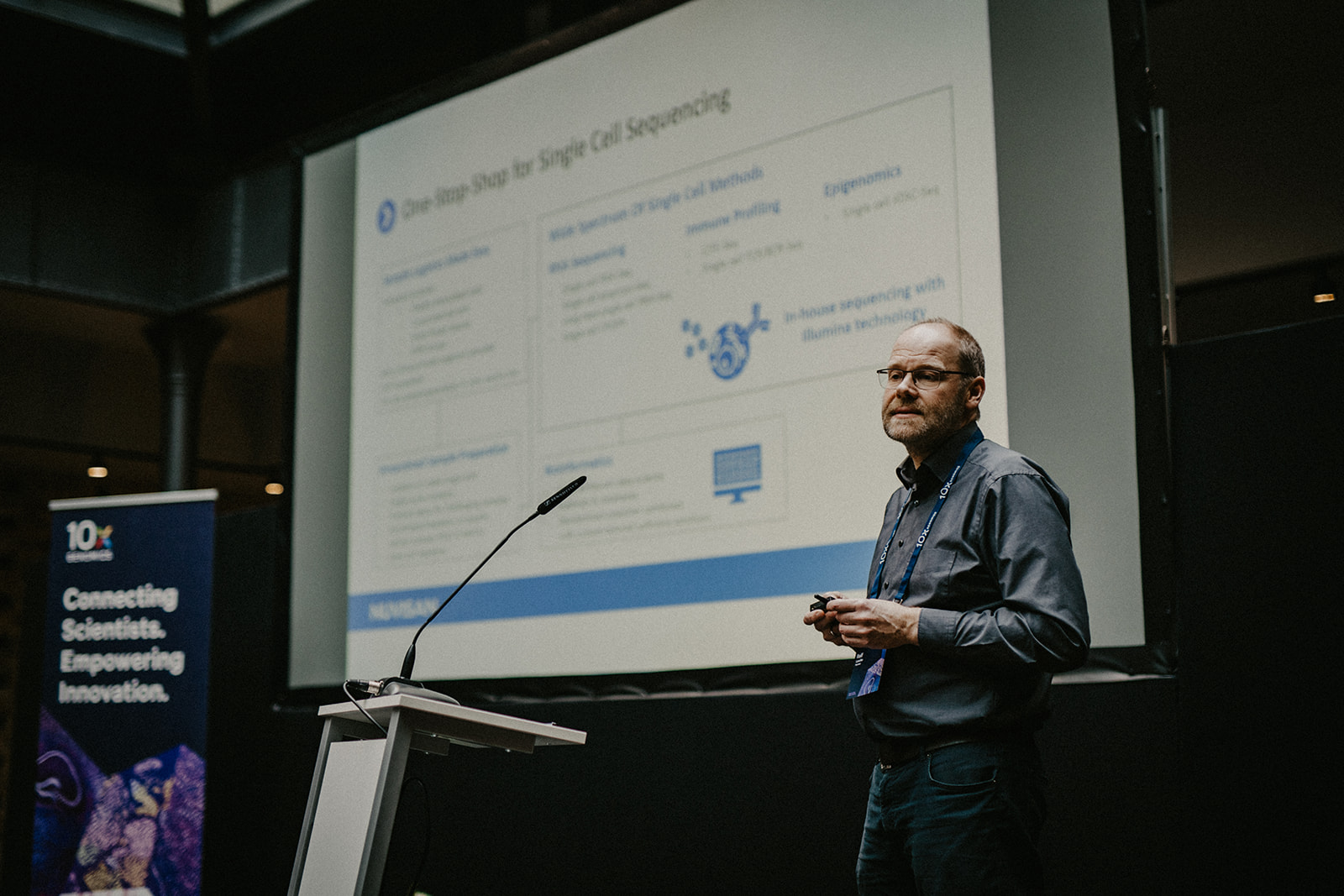
5. Ralf Lesche, PhD
Nuvisan Innovation Campus' comprehensive 10x Genomics services
At the Nuvisan Innovation Campus in Berlin, Dr. Ralf Lesche spearheads the next-generation sequencing lab, offering certified 10x Genomics services tailored for academia, pharmaceuticals, and non-profit clients. Known for their remarkable flexibility in processing various sample types, including fresh, frozen, cryopreserved, and FFPE samples, the facility is also capable of generating iPSC cells, organoids, and in vivo models on-site. With Good Clinical Practice and Biosafety Level 2–compliant facilities, Nuvisan ensures the suitability of their services for clinical samples.
Dr. Lesche's facility provides a comprehensive suite of single cell assays from 10x Genomics, along with additional options like CITE-seq, Perturb-seq, and single cell amplicon sequencing. Bioinformatics analysis is also integrated into their service portfolio. The facility's practical applications were highlighted as Dr. Lesche discussed their collaborations with a biotech company investigating gene expression in mouse lung tissue and Sanofi, focusing on post-treatment gene expression. He also showcased the core’s versatility in analyzing diverse human sample types (e.g., adipose tissue, nasal swabs, and PBMCs).
Finally, Dr. Lesche emphasized the broad applicability of 10x Genomics' single cell technologies in various projects, positioning his lab as a key player in advancing scientific endeavors, from discovery and exploratory research to drug screening and clinical trials.
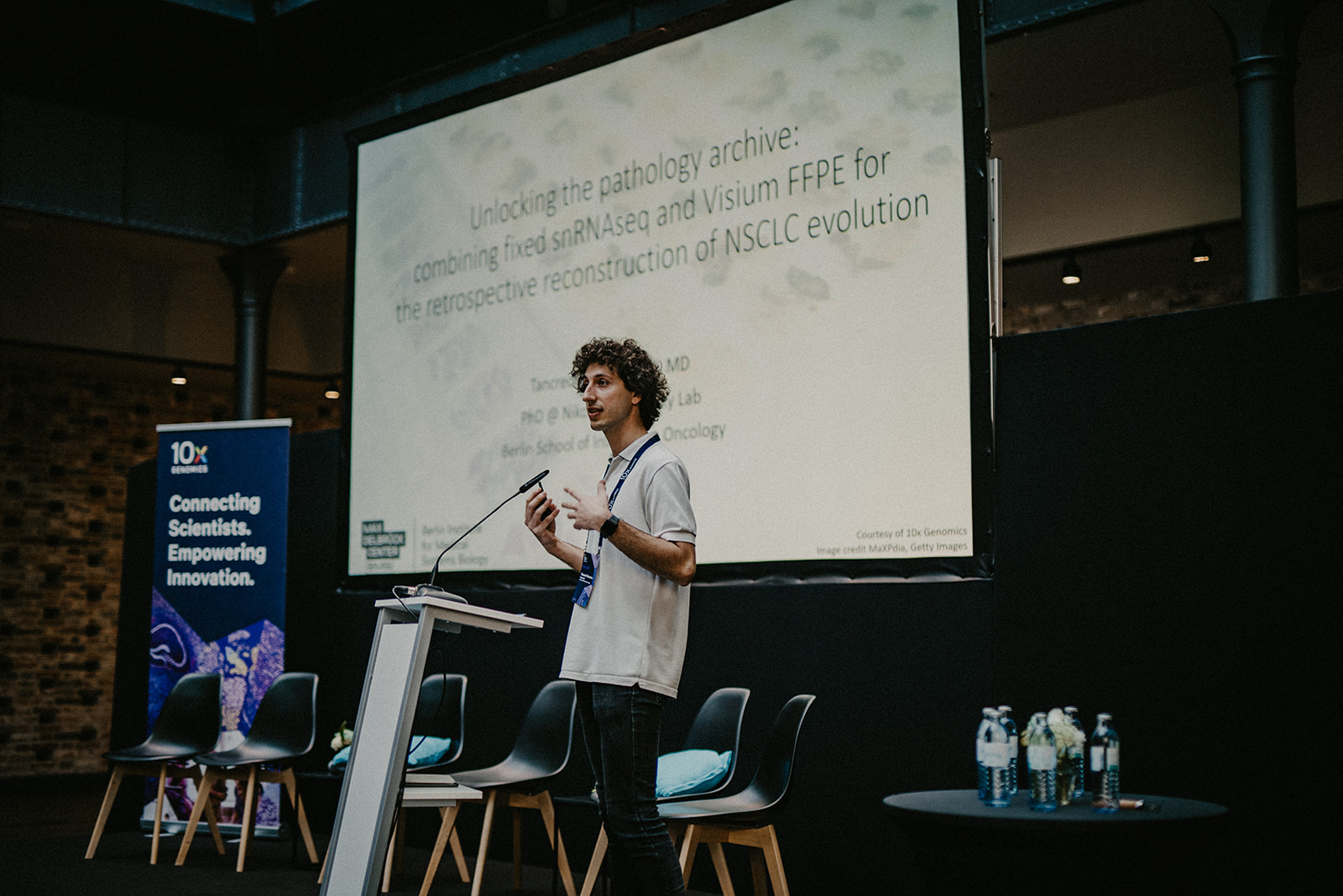
6. Tancredi Massimo Pentimalli, MD
Using 10x Genomics probe-based assays to study NSLC evolution
Dr. Tancredi Massimo Pentimalli, a computational PhD student at the Berlin Institute of Medical Systems Biology, demonstrated a pioneering proof-of-concept during his presentation at the symposium. He combined 10x Genomics kits, designed for fixed samples, to investigate the evolution of non-small cell lung cancer (NSCLC). Historically, when single cell analysis was limited to fresh samples, clinics relied on cytometry with standardized protocols. However, Pentimalli's work marks a significant shift—the availability of assays compatible with archival fixed samples is a critical aspect of his research.
Using the Flex kit, Pentimalli's team analyzed gene expression in single nuclei extracted from FFPE blocks, then employed Visium Spatial Gene Expression for FFPE on sections from the same blocks to study the spatial distribution of gene expression. Notably, they observed a surprising reduction in sequencing depth, demonstrating that 10K reads per nucleus were sufficient, challenging the conventional 30–40K reads standard. Integration with lung cancer atlas data enhanced their understanding of cell types, while the ability to infer copy number variations proved to be crucial in distinguishing normal and cancerous cells.
The team employed deep learning segmentation to identify cell types and quantify their presence in each spot on a Visium slide. This approach facilitated the creation of a spot-by-cell-type matrix, aiding in the identification of "tissue cellular niches" and "tissue niche gene pathways."
Pentimalli expanded his analysis to primary tumors, lymph nodes, and a brain metastasis, revealing insights into cell-type composition across different tumor sites. This work captured intratumor alterations, illuminated microenvironment changes, and reconstructed tumor transcriptomics over time. The Pentimalli team's innovative use of archival samples promises a profound understanding of NSCLC evolution, opening avenues for future research and therapeutic strategies.
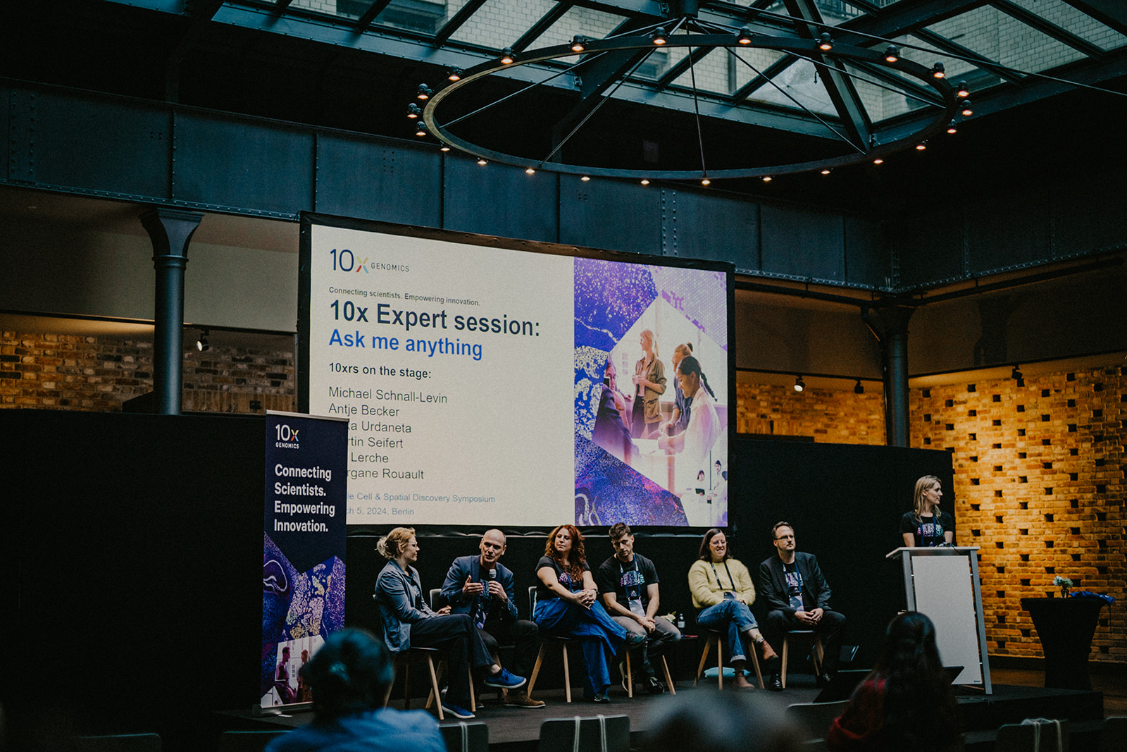
II. Requests and expectations
During the symposium, researchers expressed appreciation for 10x Genomics’ ongoing efforts to develop new kits and solutions, and also presented several requests. The users emphasized the desire for Gene Expression Flex, Visium HD, and Xenium to be compatible with species beyond humans and mice. For spatial transcriptomics technology, there was interest in incorporating proteins into the assays. Michael Schnall-Levin, PhD, Chief Technology Officer and Founding Scientist at 10x Genomics, reaffirmed this point by revealing already-in-development plans for a 20-plex protein panel on Xenium. Furthermore, researchers expressed interest in advances to the 10x Genomics’ data analysis pipelines. New ways to analyze data and collaborate using Loupe Browser were presented at the beginning of the symposium by Michael Campbell, PhD, Director of Applied Bioinformatics at 10x Genomics. Excitement surrounded upcoming products like the GEM-X product pipeline, with attendees enthusiastic about the promise of obtaining information for double the number of cells as well as potential improvements to prevent clogs and reduce sequencing costs. The anticipated Xenium 5,000-plex assay and cell segmentation kit for human and mouse samples also captured attendees' interest.
In conclusion, the symposium unveiled a wealth of information, underscoring the transformative impact of 10x Genomics technologies across diverse scientific disciplines. Researchers remain eager for further advancements, pushing the boundaries of what is possible in single cell and spatial biology research.

The symposium was also an incredible platform for collaboration and shared insights. The author, Cátia Moutinho, had the opportunity to ask attendees about the products that have most improved their research experience, as well as 10x Genomics experts and partners about tips for scientists getting started with single cell and spatial workflows.
What 10x Genomics product has made your life easier at the lab?
The attendees highlighted two products: the Flex kit and Visium CytAssist.
Tips from the field
“Be gentle with your cells while doing sample preparation.” Erika Urdaneta, Field Application Scientist at 10x Genomics
“Give yourself some time to learn when you are sectioning fresh frozen samples for spatial transcriptomics experiments.” Antje Becker, Field Application Scientist at 10x Genomics
“Pay attention to what you sequence on the same flow cell. Try to avoid sequencing short inserts with longer ones as shorter inserts tend to cluster better than longer ones.” Asija Diag, Field Application Scientist at Illumina
--- --- ---
This blog was written by guest author Cátia Moutinho, PhD. She is a scientific consultant and the founder of The Single-Cell World, a digital community of scientists dedicated to helping researchers understand all things single cell technology. You can find more of her blog articles here.