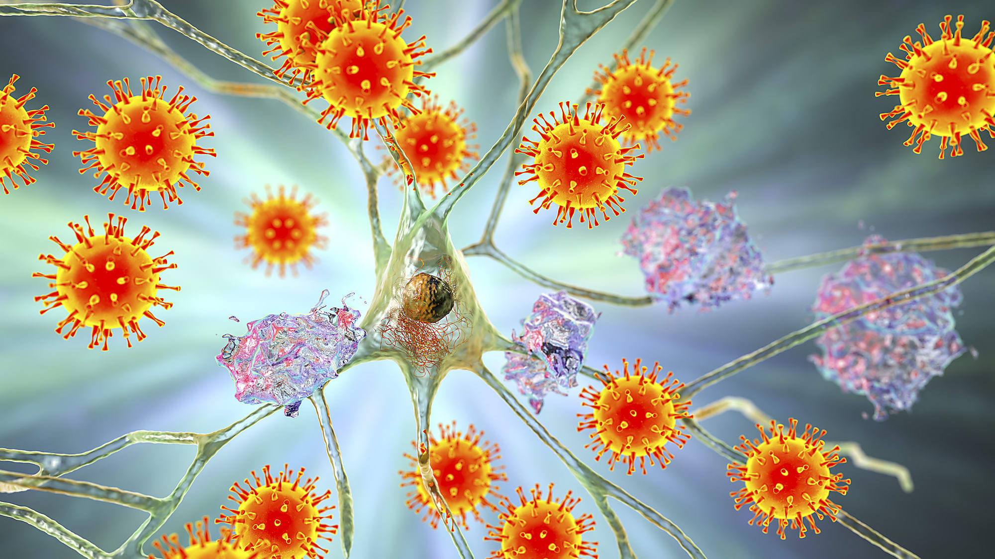
COVID and the CNS: Single cell tools shed light on the neurological consequences of COVID

From mild headaches and dizziness to potentially life-altering seizures and stroke, the impact of COVID on the brain, referred to as Neuro-COVID, is hard to ignore (1, 2). One recent survey of patients who had been hospitalized with COVID reported up to 80% of the study participants experienced at least one new neurological symptom, with 55% of them having had a clinical evaluation for the symptom (3).
Is Neuro-COVID a result of direct damage to the central nervous system (CNS)? Could a dysregulated immune response be to blame? Does it manifest as an indirect consequence of infection complications such as oxygen deprivation? These are the questions that researchers across the globe have been investigating. In this blog post, we cover some of the insights that single cell sequencing has revealed about the origins of Neuro-COVID.
A case for immune-mediated mechanisms in Neuro-COVID
Interested in how the immune system may contribute to Neuro-COVID, Heming et al. aimed to create an unbiased single cell transcriptional atlas of the cerebrospinal fluid (CSF) of these patients (4). They evaluated transcriptional profiles of cells in the CSF of patients with Neuro-COVID (eight patients). Samples from patients with viral encephalitis (VE; five patients), multiple sclerosis (MS; nine patients), and idiopathic intracranial hypertension (IIH; nine patients) were also analyzed for a total of 80,919 single cell transcriptomes generated.
Comparing the cell cluster compositions generated using the Chromium Single Cell Gene Expression assay, the authors found that the CSF of patients with Neuro-COVID contained an expanded monocyte-like cluster not seen in the IIH and VE samples. This monocyte-like cluster exhibited enhanced antigen-presenting characteristics but lowered expression of microglia gene sets, pan-monocytic markers, and border-associated macrophage markers. Thus, these cells potentially de-differentiated to a point resembling developmental macrophages.
Next, Heming et al. assessed transcriptional changes characteristic of Neuro-COVID. Interferon-driven transcripts and exhausted T-cell populations were increased in Neuro-COVID samples. However, this increased antiviral transcriptional response was not as strong as that seen in viral encephalitis (VE). When compared to VE, Neuro-COVID samples did show higher expression of genes associated with homeostatic microglia/border-associated macrophages. Further analysis indicated that severe COVID cases were correlated with a potentially impaired antiviral response compared to milder COVID cases. Lastly, T-cell receptor sequencing with the Chromium Single Cell Immune Profiling assay showed that severe Neuro-COVID was associated with a broad clonal T-cell expansion.
A case for direct damage to the brain
In the Heming et al. study, the authors highlight that “These suspected [Neuro-COVID development] mechanisms are not mutually exclusive and might coexist in individual patients.” After all, biological mechanisms are nothing if not complex. A few studies in 2020 presented evidence of detectable SARS-CoV-2 virus in some brain cells as well as activation of the brain’s immune cells, which could also be a factor in driving dangerous neuroinflammation characteristics of Neuro-Covid (5–7).
Studying the impacts of COVID on the brain, however, is complicated by the lack of available brain tissue and cell samples. Therefore, Wang et al. set out to create an in vitro model, created from human pluripotent stem cells (hPSCs) differentiated into pericyte-like cells (PLCs), that could be used to investigate SARS-CoV-2 infection of brain cells (8). Pericytes are brain cells associated with neuronal differentiation, neuroinflammation, and blood–brain barrier permeability (9), which, as demonstrated by the authors, express ACE2 RNA and protein and can be infected with SARS-CoV-2.
To create a more physiologically relevant environment to study SARS-CoV-2 infection of these cells, the researchers generated a 3D “assembloid” in which PLCs were integrated with cortical brain organoids. Once incorporated into the assembloids, the PLCs were determined to interact with astrocytes in a way closely resembling their interactions in the brain’s neurovascular unit.
The Chromium Single Cell Gene Expression assay was used to characterize these assembloids in both the presence and absence of SARS-CoV-2 infection. Interestingly, SARS-CoV-2 reads were detected largely in astrocytes of organoids only in the presence of PLCs, suggesting that astrocyte infection is mediated by PLCs. Further investigation revealed that infected astrocytes showed inflammatory and genotoxic stress responses controlled by the type I interferon pathway. These findings support the idea that viral replication hubs may exist in the brain allowing the spread of viral infections to neural cells, in other words allowing for direct damage.
Neuro-COVID research: Just the beginning
As cases of COVID continue to persist and new variants emerge, understanding the root of these neurological symptoms will be critical to helping patients recover long after the initial infection, especially since these manifestations are often linked to long COVID—health problems that present long after the initial COVID infection (10). As demonstrated in these two studies, single cell sequencing is becoming a vital tool to understand this complex process in extremely precious CNS samples.
References:
- Ellul MA, et al. Neurological associations of COVID-19. Lancet Neurol 19: 767–783 (2020). doi: 10.1016/S1474-4422(20)30221-0
- Marshall M. COVID and the brain: researchers zero in on how damage occurs. Nature 595: 484–485 (2021). doi: 10.1038/d41586-021-01693-6
- Chou SHY, et al. Global incidence of neurological manifestations among patients hospitalized with COVID-19-A report for the GCS-NeuroCOVID Consortium and the ENERGY Consortium. JAMA Netw Open 4:e2112131 (2021). doi: 10.1001/jamanetworkopen.2021.12131
- Heming M, et al. Neurological manifestations of covid-19 feature T cell exhaustion and dedifferentiated monocytes in cerebrospinal fluid. Immunity 54:164–175.e6 (2021). doi: 10.1016/j.immuni.2020.12.011
- Matschke J, et al. Neuropathology of patients with COVID-19 in Germany: a post-mortem case series. Lancet Neurol 19: 919–929 (2020). doi: 10.1016/S1474-4422(20)30308-2
- Meinhardt J, et al. Olfactory transmucosal SARS-CoV-2 invasion as a port of central nervous system entry in individuals with COVID-19. Nat Neurosci 24:168–175 (2020). doi: 10.1038/s41593-020-00758-5
- Kantonen J, et al. Neuropathologic features of four autopsied COVID-19 patients. Brain Pathol 30: 1012–1016 (2020). doi: 10.1111/bpa.12889
- Wang L, et al. A human three-dimensional neural-perivascular ‘assembloid’ promotes astrocytic development and enables modeling of SARS-CoV-2 neuropathology. Nat Med 27: 1600–1606 (2021). doi: 10.1038/s41591-021-01443-1
- Sweeney M, Ayyadurai S & Zlokovic B. Pericytes of the neurovascular unit: key functions and signaling pathways. Nat Neurosci 19, 771–783 (2016). doi: 10.1038/nn.4288
- The Lancet Neurology. Long COVID: understanding the neurological effects. Lancet Neurol 20: 247 (2021). doi: 10.1016/S1474-4422(21)00059-4