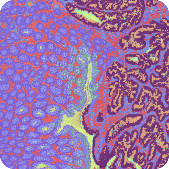
Human Prostate Cancer, Adjacent Normal Section with IF Staining (FFPE)
Spatial Gene Expression dataset analyzed using Space Ranger 1.3.0

Learn about Visium Analysis
10x Genomics obtained FFPE human prostate tissue from Indivumed Human Tissue Specimens. Original diagnosis with adenocarcinoma. The tissue was sectioned as described in Visium Spatial Gene Expression for FFPE – Tissue Preparation Guide Demonstrated Protocol (CG000408). Tissue sections of 10 µm were placed on Visium Gene Expression slides, then stained follwoing Deparaffinization, Decrosslinking, Immunofluorescence Staining & Imaging Demonstrated Protocol (CG000410).
- Block 1E333_Tp11 Section 1
- Stage II
- Total Gleason score: 7
- Sex: Male
Antibodies used for immunofluorescence (IF) staining:
- Channel 1 = 1:100 Iba1/AIF-1 (Cell Signalling, Cat. No. 36618S)
- Channel 2 = 1:100 Vimentin (Cell Signalling, Cat. No. 9856S)
- Channel 3 = 1:5000 DAPI (Thermo Scientific, Cat. No. 62248)
- Blocking buffer: 1X PBS; 2% BSA; 0.1% Tween 20; 1 U/uL Roche Protector RNase Inhibitor (based on CoA)
The IF image was acquired using Metafer Slide Scanning Microscope from MetaSystems with these settings:
- Zeiss Plan-Apochromat 20x objective
- Numerical Aperture: 0.8
- Zeiss Colibri 7 LED light source
- MetaSystems CoolCube 4c camera
Libraries were prepared following the Visium Spatial Gene Expression Reagent Kits for FFPE User Guide (CG000407 Rev A).
- Sequencing instrument: Illumina NovaSeq, flow cell H25NNDMXY (lane 1)
- Sequencing depth: 29,191 reads per spot
- Sequencing configuration: 28bp read 1 (16bp Visium spatial barcode, 12bp UMI), 50bp read2 (transcript), 10bp i7 sample barcode and 10bp i5 sample barcode.
- Dual-Index set: SI-TS-D4
- Slide: V11J26-076
- Area: D1
Key metrics were:
- Spots detected under tissue (Loupe Manual Alignment): 3,460
- Median genes per spot: 5,001
- Median UMI counts per spot: 11,444
This dataset is licensed under the Creative Commons Attribution 4.0 International (CC BY 4.0) license. 10x citation guidelines available here.