Mouse Bone Data with Custom Add-on Panel
In Situ Gene Expression dataset analyzed using Xenium Onboard Analysis 1.9.0

Learn about Xenium Analysis
Overview
Bone marrow plays a crucial role in diagnosing various conditions. By analyzing gene expression within bone marrow, researchers can gain insights into hematological disorders, immune responses, and other diseases. Bones present a unique challenge due to their hardness and inaccessibility. To address this, we have focused on optimized sample handling techniques.
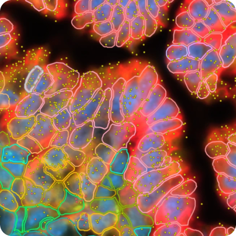
This page demonstrates Xenium In Situ Gene Expression data for mouse bone sections prepared with several decalcification methods using the Xenium Mouse Tissue Atlassing Panel with custom add-on.
How to view data
Interactively explore data with Xenium Explorer by downloading the Xenium Output Bundle (or Xenium Explorer subset) file. The subset bundle contains the experiment.xenium, morphology_mip.ome.tif, analysis_summary.html, cells.zarr.zip, cell_feature_matrix.zarr.zip, transcripts.zarr.zip, and analysis.zarr.zip files. See the Getting Started with Xenium Explorer page for more details. Follow these instructions to view the post-Xenium H&E image or image alignment file in Xenium Explorer.
Biomaterials and sample handling
Non-diseased FFPE mouse femur bone samples were obtained and processed in-house by 10x Genomics. Samples were prepared by 10x Genomics with three decalcification methods:
- With 8% formic acid (24 hours)
- With 0.5M EDTA (~3 days)
- With 15% EDTA/0.4% PFA (~3 days)
Additional guidance for bone sample preparation is available in the Analysis of Bone Tissue using Xenium v1 In Situ Gene Expression Assay Technical Note.
Tissue preparation
Decalcified tissues were prepared following the Xenium In Situ for FFPE - Tissue Preparation Handbook (CG000578). Probe hybridization, washing, ligation, and amplification were performed following the Xenium In Situ Gene Expression User Guide (CG000582).
Post-instrument processing followed the Demonstrated Protocol Xenium In Situ Gene Expression - Post-Xenium Analyzer H&E Staining (CG000613).
Gene panels
The Xenium Mouse Tissue Atlassing Panel (379 genes) was pre-designed by 10x Genomics. The panel design was informed by reference data from CZ Biohub San Francisco and The Tabula Muris Consortium and has been validated in the whole mouse pup and in nine adult tissues (liver, lung, heart, skin, kidney, spleen, colon, brain, thymus).
To enhance detection of bone cell types, we added a 100-gene custom add-on panel to supplement the identification of osteoblasts, osteocytes, osteoclasts, and chondrocytes. The add-on gene list for these datasets is available as a supplemental file. It includes panel gene name, transcript ID, and cell type annotation in CSV format.
Xenium Analyzer
The instrument run was performed following the Xenium Analyzer User Guide (CG000584). The on-instrument analysis was run with Xenium Onboard Analysis version 1.9.0.
| Metric | 8% Formic acid | 0.5M EDTA | 15% EDTA/0.4% PFA |
|---|---|---|---|
| Median transcripts per cell | 70 | 65 | 69 |
| Cells detected | 253,560 | 348,141 | 247,876 |
| Decoded transcripts per 100 µm² | 115.7 | 115.0 | 113.0 |
| Total high quality decoded transcripts | 24,531,426 | 40,352,654 | 27,864,048 |
| Region area (µm²) | 51,981,815 | 75,035,166 | 55,0003,268 |
This dataset is licensed under the Creative Commons Attribution 4.0 International (CC BY 4.0) license. 10x citation guidelines available here.