
Atera World Tour 2026
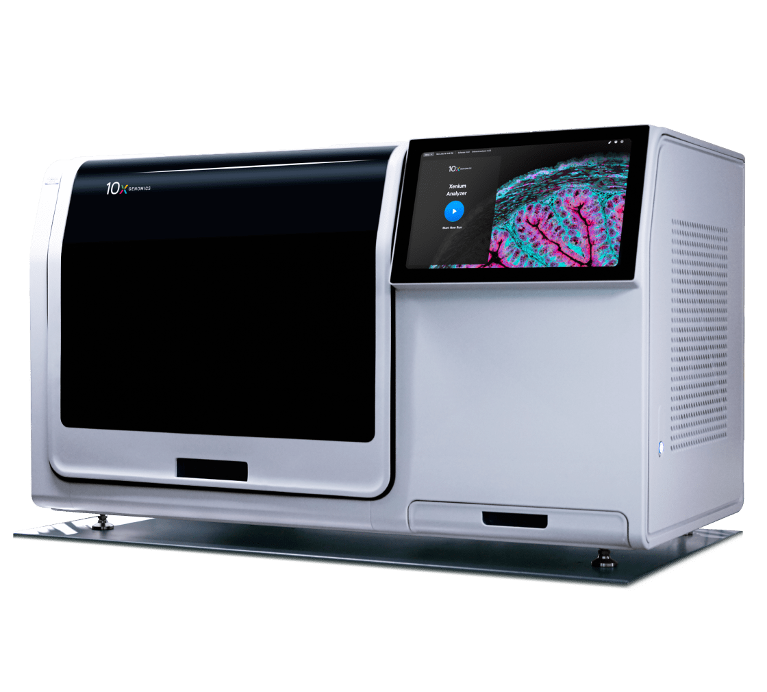
Find a stop near youA state-of-the-art single cell spatial imaging platform
Built to deliver exceptional image and data quality
Xenium offers industry-leading speed and throughput
Using multimodal cell segmentation, analyze 472 mm² of tissue per run, targeting up to 480 genes in < 3 days or up to 5,000 genes in < 6 days.
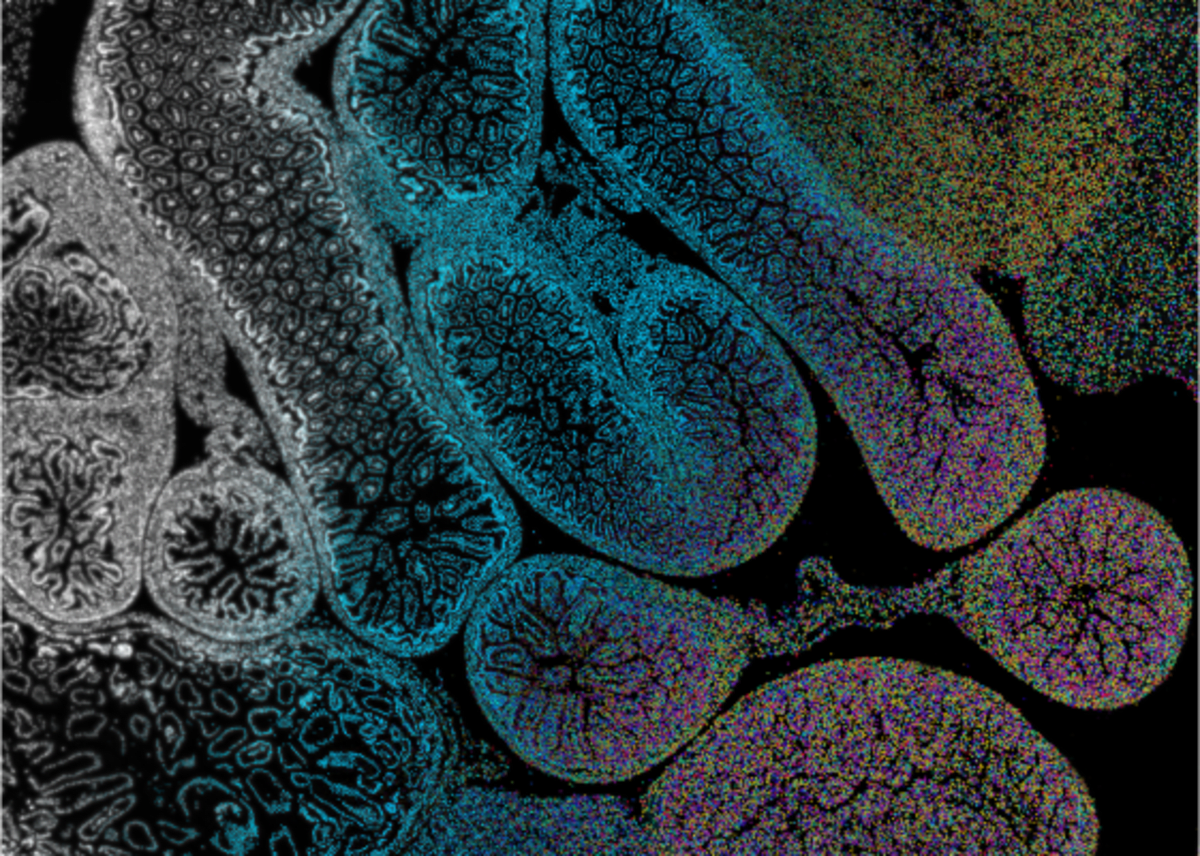
Converts terabytes of 3D imaging data into biologically meaningful insights
One-of-a-kind onboard analysis software performs high-powered computation on-instrument. Internal image sensor data is converted into ready-to-explore formats as soon your run finishes.
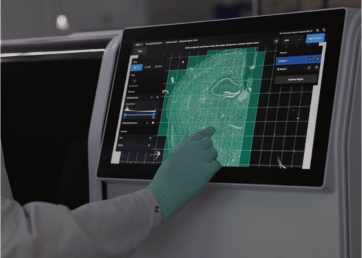
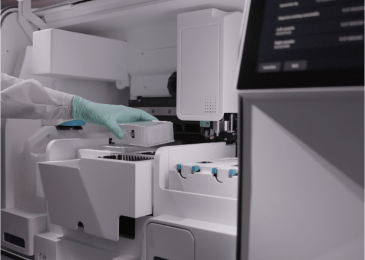
Delivers an unparalleled user experience
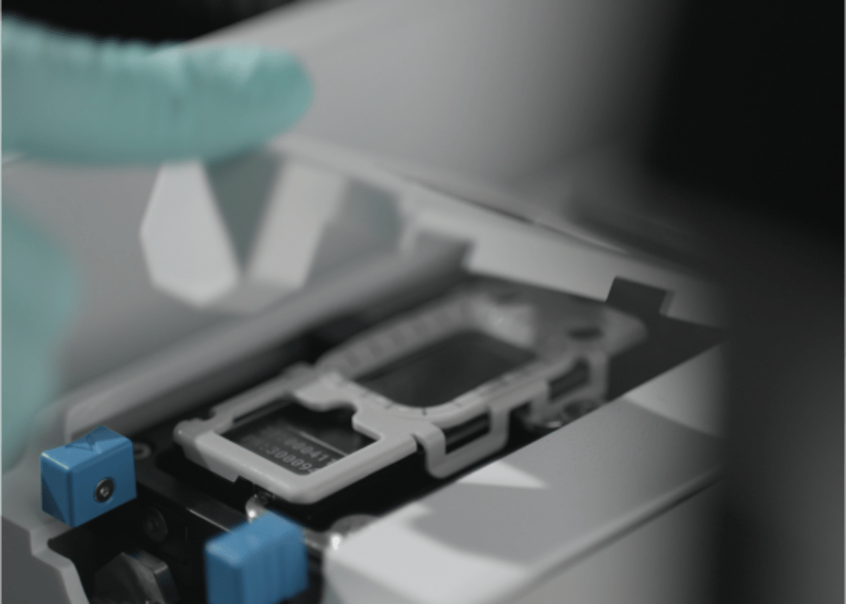
Simple run setup and cleanup operations, an intuitive touchscreen, and ergonomic design make instrument operation a breeze.
Expands with upcoming innovations
Flexible fluidic architecture allows the Xenium Analyzer to leverage new functionalities, assay enhancements, and more.
The ultimate single cell spatial biology experience
| Instrument | 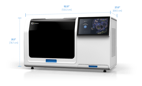 Xenium Analyzer |
| Dimensions (W x D x H) | 52.5” x 27” x 31” (59” with door open)
133.3 cm x 68.5 cm x 78.7 cm (149.8 cm with door open) |
| Resolution | Transcript XY-localization precision < 30 nm and Z-localization precision < 150 nm
Pixel size = ~0.2 µm/pixel |
| Number of slides per run | 2 slides |
| Throughput (area per time) | 472 mm² of tissue per run when targeting up to 480 genes in < 3 days or up to 5,000 genes in < 6 days*Explore Runtime Planner |
| Total tissue area analyzable per run | ~ 472 mm² |
| Sample prep | 2–3 days sample prep with 4–6 hrs of hands-on time |
| Data output file formats | Cell-feature matrices: MEX, HDF5, Zarr
Transcripts and segmentation boundaries: CSV, Parquet, Zarr
Tissue images: OME-TIFF |
| Connectivity | Compatible with 10x Genomics Cloud instrument management |
Explore the science behind Xenium
See what Xenium users are saying
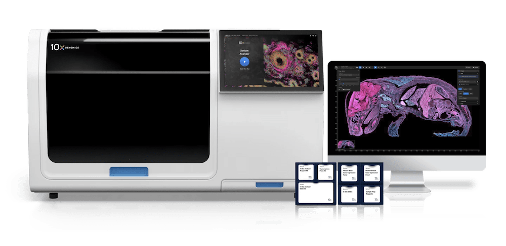
Xenium In Situ Platform
Building on our years of innovation in single cell and spatial technologies, Xenium was designed to transform the way researchers can characterize RNA, protein, and cells in their native tissue environment.